Figure 7.
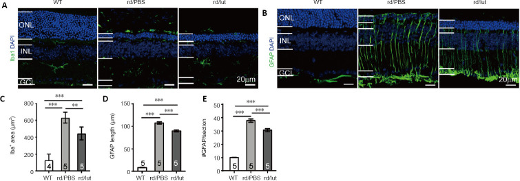
Luteolin (lut) inhibites reactive gliosis in the retinas of rd10 mice.
(A, B) Retinal slices stained for DAPI (blue) and Iba1 (A) or GFAP (B) (green: Alexa Fluor-488) at P26. In the retinas of rd10 mice (rd), Iba1-positive microglia were present throughout the outer retina and GFAP was highly expressed in Müller cells distributed throughout the whole retina. Luteolin decreased both Iba1 and GFAP staining in the outer retina. Scale bars: 20 μm. (C–E) Area of Iba1-positive staining (C), length of GFAP-positive processes (D) and number of GFAP-positive processes (E) per image (320 μm × 320 μm) for each group. Data are expressed as mean ± SEM. **P < 0.01, ***P < 0.001 (one-way analysis of variance followed by Tukey’s post hoc test). Numbers within/near bars indicate quantities of mice tested. DAPI: 4′,6-Diamidino-2-phenylindole; GCL: ganglion cell layer; GFAP: glial fibrillary acidic protein; Iba1: ionized calcium-binding adapter molecule 1; INL: inner nuclear layer; ONL: outer nuclear layer; P: postnatal day; PBS: phosphate-buffered saline; WT: wild-type.

