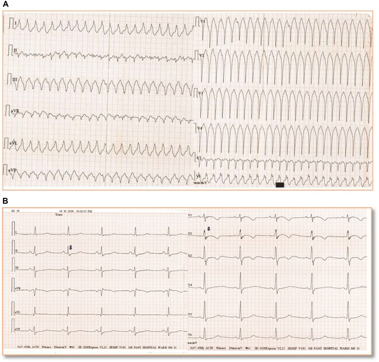
Figure 1.
(A) Twelve-lead electrocardiogram (at 25 mm/s speed) recording of monomorphic ventricular tachycardia episode recorded at time of presentation to emergency—left axis deviation, QRS duration in lead I >120 ms, QRS transition in V6, notching of QRS in V6 favour a diagnosis ventricular tachycardia of right ventricle origin. (B) Electrocardiogram in sinus rhythm after opening the 40 Hz filter at double amplification and 50 mm/s speed recording. Epsilon waves in leads V1–V3 (blue arrows) with T-wave inversion and terminal activation duration of 60 ms. Epsilon waves are also noted in leads II, III, avF (blue arrows) that suggests concomitant left ventricular involvement.

