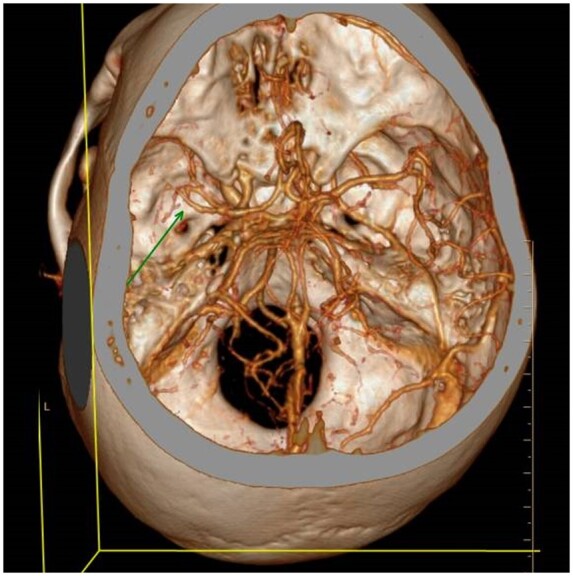
Figure 3.

Reconstructed image from brain computed tomography angiography demonstrating changes in the left hemispheric blood vessels middle cerebral artery and anterior cerebral artery A1 segment correlating with cerebral vasculitis or angiitis. Arrowhead—narrow, irregular, and beading like appearance of middle cerebral artery M1, M2 segments.
