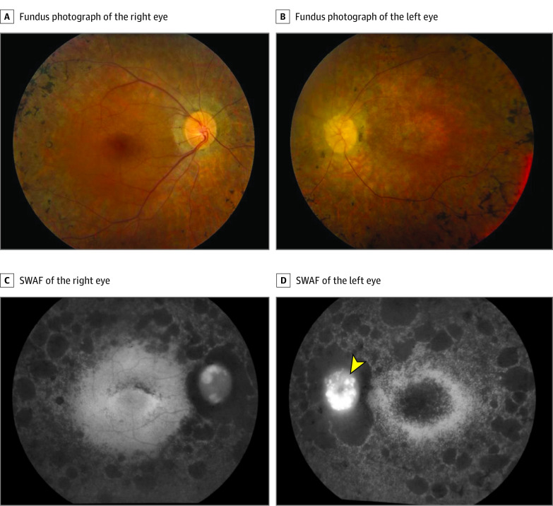
Figure 1. In Vivo Imaging of the Patient Shows Typical Signs of Retinitis Pigmentosa, With Degenerative Changes More Advanced in the Right Eye Compared With the Left Eye.
Fundus photography and short-wavelength fundus autofluorescence (SWAF) show severe retinal vascular attenuation, optic disc pallor, and retinal atrophy with bone spicules in both eyes. The fovea is globally hypoautofluorescent in the left eye (D), whereas there are speckled patches of hypofluoresence in an arc superior to the fovea in the right eye (C).

