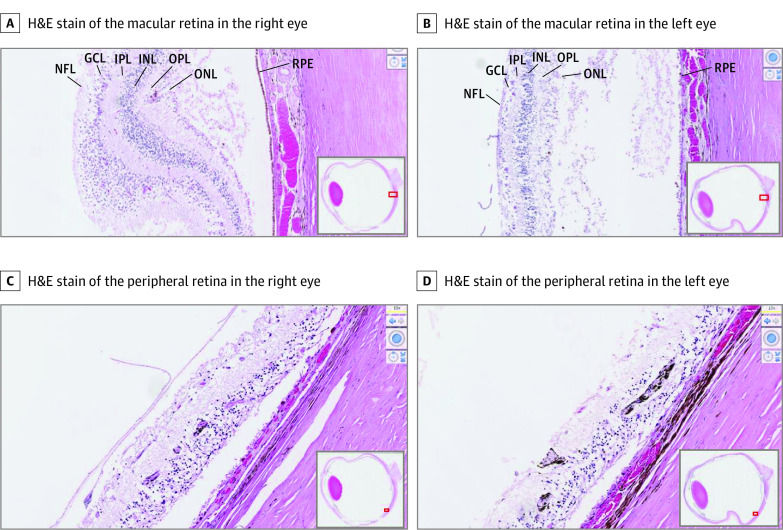
Figure 2. In Vitro Histopathological Studies Confirm More Advanced Degeneration of Retina in the Left Eye Than the Right Eye.
Representative hematoxylin-eosin (H&E)–stained histology of the right and left eyes taken at ×100 original magnification shows loss of photoreceptors and attenuation of layers of the retina to be more advanced in the left eye, continuing to the extent of the ganglion cell layer (GCL). This is evident both in the central (A and B) and peripheral (C and D) retina. Along with retinal atrophy, retinal pigment deposits are particularly evident in the peripheral retina in the left eye. INL indicates inner nuclear layer; IPL, inner plexiform layer; NFL, nerve fiber layer; ONL, outer nuclear layer; OPL, outer plexiform layer; RPE, retinal pigment epithelium.

