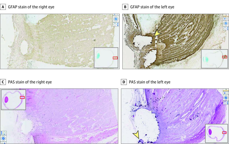
Figure 3. In Vitro Histopathological Studies Demonstrate Greater Optic Disc Drusen in the Left Eye Than the Right Eye.
Images were taken at a magnification of ×25. Additional images of right and left eyes reveal artifact of where optic disc drusen once were. Glial fibrillary acidic protein (GFAP) staining shows gliosis at the optic nerve head (B), a corollary of the extent of retinal atrophy. There appear to be fragments of positive periodic acid–Schiff (PAS)–stained material at the edge of the gap in panel D (arrowhead), representative of remaining drusen.

