



Abstract
Endocardial fibroelastosis (EFE) signifies the pathological process by which collagen and elastin are focally or diffuse deposited in the endocardium of the left ventricle. The new layer causes left ventricular dysfunction sometimes with fulminant progression to heart failure. EFE is a major component in many congenital heart abnormalities but can also occur in the absence of heart malformations, either as a primary process or in response to cardiac injury. The endothelial–mesenchymal transition (EndMT) abnormalities seem to be main pathogenic factor in fibroelastosis development. The “gold standard” for diagnosis of primary EFE (pEFE) is the histological examination. Additionally, genetic studies may help to establish the natural course of the disease and to communicate prophylactic measures to family members of the affected child. Moreover, in the newborn, EFE takes the form of dilated cardiomyopathy (DCM) with unfavorable evolution. The proper management should be established considering negative prognostic factors, involving early transplantation, drug therapy and long-term follow-up.
Keywords: endocardial fibroelastosis, dilated cardiomyopathy, mutations, gene therapy
⧉ Introduction
Endocardial fibroelastosis (EFE) is a complex process consisting in collagen and elastin deposits in the endocardial layer of the heart [1]. The accumulation of inelastic collagen fibers and elastin is made by fibroblasts, that in turn generate acellular fibrocartilaginous extracellular matrix, responsible for diffuse thickening of the endocardium. This accumulation of collagen and elastin determine the overgrowth of fibrous tissues leading to important morpho-functional changes in the left ventricular endocardium, particularly in the first year of life in infants and children.
EFE is considered to arise either de novo with no evident reason, either involving a genetic background with two forms, namely X-linked recessive and autosomal recessive patterns in the context of genetic mutation [2]. Also, infection and viral etiology were taken into consideration.
EFE can be primary or secondary, each form of disease having a different mode of evolution [1]. While primary fibroelastosis is not related to cardiac anomalies, secondary type seems to be associated with congenital heart malformations as hypoplastic left heart syndrome (HLHS) or left ventricular non-compaction (LVNC). The late disorder is determined by the disruption of blood flow in the context of an incompetent left ventricle, to which is added an abnormal ejection tract [3,4]. Flow disturbances can be avoided by inhibition of formation of EFE tissues. So, the effective measures are still needed to preserve the ventricular diastolic compliance and renew myocardial growth [4].
Aim
This review aims to outline the epidemiology of EFE in children, its outcomes, etiology, genetic and morphological changes, clinical aspects in particular of primary EFE (pEFE) and to offer a depth approach to diagnosis, focusing on histological examination and genetic studies. In addition, we discuss the importance of echocardiography or computed tomography (CT) criteria associated with the fetal cardiovascular score in order to identify this life-threatening condition.
⧉ Pathogenic overview
The endothelial–mesenchymal transition (EndMT) represents a process that normally takes places in cardiac embryogenesis, through which epicardial cells become fibroblasts. In the neonatal period, abnormalities of this phenomenon initiate the fibroelastosis development. Studies of gene expression in this cell population have identified an increased level of transforming growth factor beta (TGFβ). Furthermore, administration of bone morphogenetic protein 7 (BMP7), an antagonist of the TGFβ signaling pathway, resulted in regression of the fibrous area and inhibited the accumulation of fibroblasts [2, 5].
Studies performed on resected cardiac tissue affected by anomalies, such as left hypoplasia, have revealed the involvement of the endocardium in initiating and supporting pEFE, through the same process of EndMT. The administration of TGFβ inhibitors was successful in this context as well. All this data suggests a different origin of fibroblasts depending on the type of pEFE [5].
Researchers tried to establish echocardiographic criteria for diagnosis and staging of pEFE in both prenatal and postnatal periods, especially in the case of its association with aortic stenosis. The aim was to determine the prognosis, which depends on the success rate of surgical procedure that dilates the stenosis in uterus or the probability of normal evolution of a hypoplastic left ventricle after surgical resection of the area of fibroelastosis [6]. Four stages of prenatally assessed pEFE severity were proposed, starting from 0 – absent, 1 – mild (spots of left ventricular hyperechogenicity, including papillary muscles), 2 – moderate (non-confluent hyperechogenic areas), 3 – severe (linear hyperechogenicity). The severity of fibroelastosis correlated with geometric and functional aspects of the left ventricle and postnatal prognosis after in utero surgery [6,7]. pEFE grading together with fetal cardiovascular score allow a proper management of the problematic pregnancies [7].
Corroborating evidence from echocardiographic and pathological examination led Seki et al. [8] to conclude that pEFE has pathological characteristics distinct from those of dilated cardiomyopathy (DCM). The idea was supported by studies on tissue samples and imaging data from a cohort of 52 patients of three months to 17 years aged who had undergone a heart transplant for DCM. The authors considered that 14 patients presented typical features of pEFE, as follows: diffuse thickening of the left ventricular endocardium, elevation of papillary muscles, thickening of the free edge of the mitral valve and specific aspect of the organization of elastic fibers at pathological examination [8].
Multiple mechanisms of pEFE development are discussed, including obstruction of subendocardial lymphatic drainage, blockage of the smallest cardiac veins (Thebesian veins), and premature closure of the foramen ovale [2]. pEFE tends to manifest in the first year of life, most often having a sudden onset, precipitated by a respiratory infection. Many of the cases of idiopathic DCMs are considered to be, in fact, pEFE. The clinical examination reveals tachypnea, fatigue, decreased appetite, diminished apex shock, deafening heart sounds, and ventricular gallop. Signs of heart failure may be present as well. The electrocardiogram detects left ventricular hypertrophy, isoelectric or negative T waves in precordial leads, R wave >20 mm in V6, S >20 mm in V1, normal or discrete left QRS axis [5].
Treatment consists in administering Digoxin in 60–80 μg/kg loading dose, followed by 15–25 μg/kg along with diuretics and oxygen if needed. Particular cases are those of pEFE occurring in newborns with mothers suffering from autoimmune diseases. In these cases, the administration of intravenous immunoglobulin G (IgG) together with glucocorticoids and angiotensin-converting enzyme inhibitors proved favorable results [5, 7].
DCM is a chronic heart condition, a phenotypic expression of the interaction between multiple genetic and non-genetic factors. It is defined by the presence of left ventricular dilation and contractile dysfunction, in the absence of hemodynamic disturbances or coronary heart disease [9]. It is one of the most common causes of heart transplantation worldwide, counting over 50% of heart transplant cases in children aged between 1–10 years [8].
DCM can be caused by mutations of the genes involved in the coding of sarcomeric, cytoskeletal, and nuclear envelope proteins. In such cases, DCM can occur in a pattern of neuromuscular manifestations, encouraging family screening. Additionally, it can be the result of an inflammatory process most often caused by cardiotropic viral infections or an adverse drug reaction. Moreover, the endocrine or autoimmune diseases, consumption of toxic substances such as ethanol, cocaine and amphetamines may also lead to DCM. Rare forms are peripartum, Takotsubo, and tachycardia-induced cardiomyopathy [10].
Echocardiographic diagnostic criteria for DCM are represented by shortening fraction <25% [>2 standard deviation (SD)] or ejection fraction <45% (>2 SD) and left ventricular telediastolic diameter >117% (>2 SD from age-adjusted predicted value and body surface) [10,11].
Studies regarding the association between EFE and DCM revealed mutations affecting the nexilin F-actin binding protein (NEXN) and nebulette (NEBL) genes, encoding a protein involved in postnatal T-tube formation, and a specific cardiac protein involved in the tropomyosin–troponin interaction [12,13]. In the case of NEBL, screening of patients with DCM and pEFE phenotype led to the identification of gene mutations that induced the same phenotype when recreated in murine specimens [13]. Murine models in which both NEXN alleles were experimentally inactivated died shortly after birth, developing DCM. The anatomopathological examination of the cardiac tissue in these cases displayed features of EFE [14].
⧉ Key players in EFE assessment
Histological studies are the “gold standard” to understand the mechanisms of fibroelastosis. Lurie et al. concluded that both infants and young children can benefit for transvascular endomyocardial biopsy, facilitating evaluation of early active fibroelastosis [15].
The normal endocardium has three sublayers: (i) endothelium which consist of simple squamous epithelium and subendothelial connective tissue (inner portion that lines all heart cavities); (ii) fibroelastic connective tissue that intertwined with thin smooth muscle cells (middle layer); (iii) subendocardial layer joining the endocardium to myocardium by loose connective tissue (deeper layer) [16].
The smooth muscle cells layer is predominantly involved in the progressive process of EFE [17]. Electron microscopy revealed the chronological sequence of changes that occur in a heart affected by EFE. The most important steps are: (i) hyperplasia of smooth muscle cells; (ii) translocation in the juxtamyocardial layers; (iii) transformation into leiomyoid cells (features of smooth muscle cells and fibroblasts). Therefore, the elastic and collagen fibers seem to be secreted mostly by leiomyoid cells [17]. This process can be triggered by various factors; therefore, some authors consider the term of idiopathic EFE to be more appropriate. The first identified damaging factors for EFE are Coxsackie and mumps virus, genetic syndromes, autoimmune disease, metabolic and vascular disorders [18,19].
The finding that various pathogens found in the maternal blood can trigger a cardiac response in the fetus, opened the way for studies of fetal immune response at myocardium level. Patients born from mothers with anti-Ro or anti-La antibodies develop fibroelastosis associated with different types of cardiomyopathies. Their myocardium specimens were found to have increased IgG, IgM and T-lymphocytes [19].
Fernandes et al. used autopsy specimens to study the different impact of immune cells in the pathogenesis of (i) fetuses with EFE and DCM; (ii) fetuses with secondary EFE (caused by outflow tract obstruction); and (iii) fetuses with normal hearts (control group) [19]. In their study, they excluded (a) syndromic cases; (b) intrauterine infection and/or maternal autoimmune history; (c) hypothyroidism; and (d) prenatal drug abuse. The results showed that EFE/DCM group had significantly fewer B-cells and macrophages and higher cluster of differentiation (CD)4/CD8 ratio compared to the control group. The secondary fibroelastosis group had lower T-lymphocytes than the normal control group. These findings indicate either an initial shortage of immune cells in the myocardium, either their depletion [19]. In both situations, it seems that the myocardium is unable to fight against infections in the context of aggressive inflammatory process. Such theories form the basis for immune therapies in secondary EFE.
⧉ Evidence for genetic and morphological crosstalk in EFE
For the idiopathic cases, further research into the underlying causes is needed to highlight the genetic aspects of EFE, revealing specific mutations that determine specific histopathological findings in the affected heart. Table 1 summarizes the main genes involved in the development of fibroelastosis, their chromosomal position, the protein encoded by each of them and the role those proteins have, as found in the literature.
Table 1.
Genetic mutations associated with fibroelastosis
|
Affected gene / gene location |
Protein role |
Phenotype |
Heart histopathology |
|
TAZ / Xq28 |
Transacylase involved in mitochondrial cardiolipin maturation [20]. |
DCM+EFE; proximal myopathy; growth retardation; organic aciduria; neutropenia [20,21]. |
Myocardial fibrosis and cardiomyocyte apoptosis; sarcomere Z-lines normal morphology; A-bands and M-lines were not delineated poorly delineated; disorganized mitochondrial cristae [21]. |
|
CSRP3 / 11p15 |
Muscle-specific LIM-protein; myogenesis and sarcomere assembly [22]. |
Fatigue; DCM+EFE; congestive heart failure; defective neuromuscular transmission [22]. |
Disruption of cardiomyocyte cytoarchitecture; irregularly arranged myofibrils [22]. |
|
ACTN2 / 1q43 |
Cytoskeletal protein localized in the Z-disc [23]. |
DCM; LVNC; congenital myopathy [23]. |
Diffuse fibrosis; diminished signal for plakoglobin [23]. |
|
NEXN / 1p31.1 |
Rapidly progressive DCM; Diminished cardiac contractility [14, 24]. |
Collagen-positive mural masses; absence of elastic fibers and presence of fibrin; disrupted connections between sarcoplasmic reticulum and sarcolemma; absent sarcolemmal invaginations [14, 24]. |
|
|
NEBL / 10p12.31 |
Endocardial thickening; deposition of elastic tissue and collagen; progressive Z-line abnormalities; enlarged and deformed mitochondria; abnormal lysosomes and mitochondrial remnants; accumulation of lipids in cardiomyocytes [13, 25]. |
||
|
LDB3 / 10q23.2 |
Cytoskeletal protein binding and muscle alpha-actinin binding [26]. |
LVNC; DCM-association with mitral valve prolapse; myofibrillar myopathy [26]. |
Z-disk streaming and disintegration; disorganized sarcomeres and myofibrils; aggregates may contain myotilin, desmin, αB-crystallin, dystrophin [26]. |
|
PRDM16 / 1p36.3 |
Binds DNA and functions as a transcriptional regulator; functions as a repressor of TGFβ signaling [27]. |
LVNC; EFE; DCM; MDS; AML [27]. |
Myocardial thickening with a compacted and non-compacted area; myocyte disarray with staghorn-like spaces, in the non-compacted layer [27]. |
ACTN2: Actinin alpha 2; AML: Acute myeloid leukemia; CSRP3: Cysteine and glycine rich protein 3; DCM: Dilated cardiomyopathy; DNA: Deoxyribonucleic acid; EFE: Endocardial fibroelastosis; HCM: Hypertrophic cardiomyopathy; LDB3: LIM domain binding 3; LVNC: Left ventricular non-compaction; MDS: Myelodysplastic syndrome; NEBL: Nebulette; NEXN: Nexilin F-actin binding protein; PRDM16: Positive regulatory domain 16; TAZ: Tafazzin; TGFβ: Transforming growth factor beta.
⧉ Diagnosis approach in EFE
EFE usually manifests in the first month of life. While the secondary form is related to other cardiac abnormalities or systemic diseases, the primary form is often confused with idiopathic DCM [28].
As stated before, the “gold standard” for pEFE diagnosing is the histological examination, followed by genetic studies that may help to establish the natural course of the disease and to communicate prophylactic measures to family members of the affected child.
Cardiac CT identifies aspects of apical calcifications in fibroelastosis [29], while magnetic resonance imaging (MRI) allows the detection of cardiac fibrosis, assessment of ventricular wall thickness, quantification of overall ventricular function and regional contractility, intracavitary thrombus formation, all known as important prognostic factors [30].
Echocardiography and electrocardiography (ECG) are useful in differential diagnosis, but MRI and/or CT in conjunction with the typical features of microscopic examination provide vital information. pEFE may present in the form of premature ventricular contractions and non-sustained ventricular tachycardia that may easily change into life-threatening conditions that require fast identification and prompt intervention [31].
The literature describes cases of children having conduction abnormalities or frequent premature ventricular contractions. While initial ECGs and echocardiographic aspects seem to be normal, recurrent examinations and detailed investigations helped reveal the true nature of the underlying disease as EFE, allowing successful treatment courses [32].
Ino et al. conducted a retrospective study on 52 patients aged eight months to two years diagnosed with fibroelastosis, assessing the specificity and sensitivity of clinical and echocardiographic criteria and negative prognostic factors [33]. The major clinical signs were dyspnea, gastrointestinal disorders, fever, weight loss and hypotonic neurological disorders. Clinical examination revealed systolic murmur of mitral regurgitation and ventricular galloping sounds. ECG signs of left ventricular hypertrophy were present, with ST segment and T-wave inversion in V5 and V6, as well as left atrial or biatrial enlargement. An unfavorable prognosis was found among patients with persistent ECG changes despite appropriate drug therapy, associated with an ejection fraction less than 33% and a cardiac index of less than 3.5 L/min/m2. In this group, mortality at four years was 100%, suggesting that the identification of the previously mentioned negative prognostic factors would be an indicator for early heart transplantation [33]. In the group with a four-year follow-up, the persistence of electrocardiographic abnormalities was associated with an increased risk of death, suggesting the need for long-term patient monitoring [33].
In our experience, we diagnosed one case of fibroelastosis in a 7-month-old female patient, born naturally, at 40 weeks of pregnancy, weighing 3150 g at birth. The child was transferred to our Unit for the treatment of an interstitial pneumonia with unfavorable evolution under antibiotic treatment. Clinical examination at admission revealed expiratory dyspnea, thoraco-abdominal asynchrony, abnormal, piston-like movements of the head, perioral cyanosis and pale skin, respiratory rate of 50 breaths/min, coarse vesicular murmur. The heart sounds were rhythmic, with no added murmurs, with a heart rate of 190 beats per minute (bpm) and blood pressure of 91/71 mmHg. The ECG revealed sinus tachycardia, with a frequency of 200 bpm, QRS axis at 65 degrees, P wave axis at 60 degrees, duration PR/QRS/QTc: 0.12/0.08/0.40 s, aspect of hypertrophy left ventricular based on voltage criteria [34] and left atrial hypertrophy. Echocardiographically, the appearance of subendocardial fibroelastosis was detected, with a dilated left ventricle (Figure 1), ejection fraction of 10% and a shortening fraction of 4%, tricuspid and mitral reflux grade II (Figure 2), and minimal pericardial reaction.
Figure 1.

Long parasternal view. Dilated left ventricle
Figure 2.
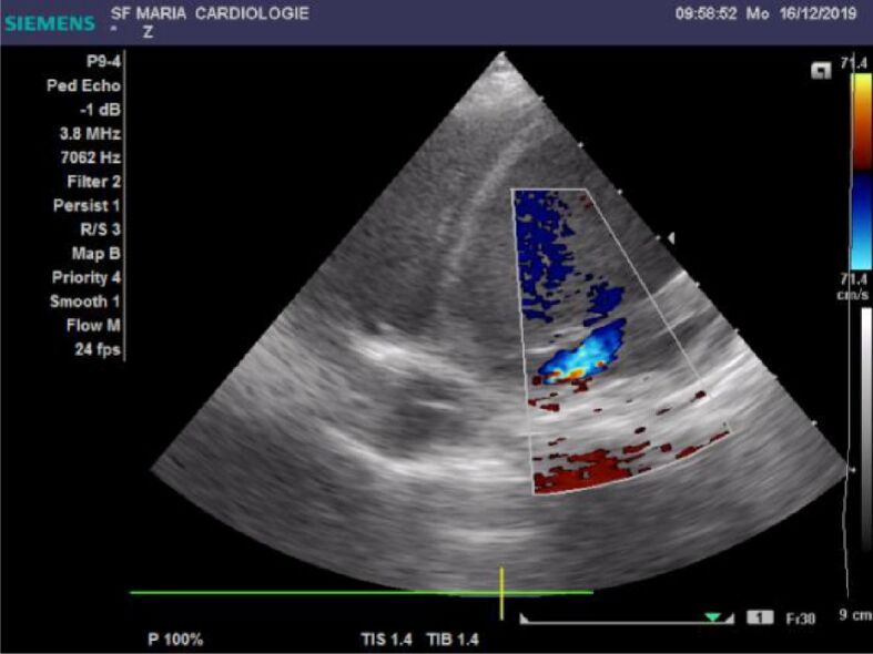
Apical four chamber view. Mitral insufficiency and hyperechoic endocardium
The patient was admitted to the Intensive Care Unit and treated with Digoxin 0.15 mg intravenously (iv) bolus then 0.075 mg iv, Furosemide 5 mg every 12 hours, along with Acetylcysteine 100 mg per day and Dexamethasone 1 mL every 12 hours. Initially, the patient maintained peripheral oxygen saturation over 97%, while still tachycardic and polypneic. Twelve hours after presentation, she developed fever (38.5°C), with cold and marbled extremities, blood pressure of 77/35 mmHg, and respiratory rate of 50 breaths/min. She was intubated and mechanically ventilated. The patient had a cardiorespiratory arrest with initial response to resuscitation, but whose recurrence led to death. At autopsy, gross morphological examination described cardiomegaly caused by fibroelastosis, with thickened endocardial deposits, proximal insertion of the papillary muscles, damage to the edge of the mitral valve (Figure 3).
Figure 3.

Macroscopic heart feature: ventricular and atrial fibrous thickening of endocardium
Microscopically, aspects of myocytolysis in the sub-endocardial area have been described along with important fibroelastic tissue in endocardium and subendocardium (Figures 4,5,6,7). Our patient was in the category of cases with a severe prognostic, as evidenced by ultrasound and clinical criteria of severity, which resulted in a rapid death, 15 hours after the initial presentation. The family history did not reveal the existence of other cases of fibroelastosis in the family and did not raise the suspicion of a hereditary syndromic pathology. However, this line of investigation needs to be followed in any type of cardiomyopathy, including in those related to fibroelastosis.
Figure 4.

Abundant fibroelastic tissue in subendocardial layer and myocardium with vascular congestion (HE staining, ×400). HE: Hematoxylin–Eosin
Figure 5.

Fibrous thickening of endocardium associated vacuolar change secondary to myocytolysis (HE staining, ×100)
Figure 6.

Masson’s staining (×200) shows increased collagen deposits in subendocardial area
Figure 7.

Important fibrous thickening of subendocardial layer – detail (Masson’s staining, ×400)
As studies into the pathogenicity of EFE continue, more dilemmas are born. The distinction between primary and secondary fibroelastosis as well as the classification of this disease amongst other cardiomyopathies, is reason for creating two sides. Some studies consider fibroelastosis as a reaction of the myocardium to various injuries, rendering the term pEFE as inappropriate, while other consider pEFE as a self-standing disease, which arises because of genetic and epigenetic interferences.
Whatever the results of this debate are, it is for the moment clear that EFE/pEFE leads to DCM-like phenotypes more often than to a restrictive form of cardiomyopathies. This observation has important therapeutical implications. Regenerative therapy as treatment for severe heart failure has gain a lot of attention in the past decade, cardiac reprogramming by means of transplantation of induced pluripotent stem cell (iPSC)-derived cardiomyocytes being also investigated for cases with DCM [35]. Ieda investigated Sendai virus (SeV) and adeno-associated viruses as vectors for delivering human cardiac reprogramming factors to transform cardiac fibroblasts into induced cardiomyocyte-like cells (iCMs) [35]. Whether this attempted remediation is feasible for EFE cases is an exciting research direction.
⧉ Conclusions
Fibroelastosis is a rare condition with a relatively poor prognosis. The suspicion of fibroelastosis can rise from the prenatal period and can be managed using echocardiographic criteria associated with the fetal cardiovascular score. Early detection allows the initiation of neonatal treatment as soon as possible, thus preventing further complications. The proper management should be established considering negative prognostic factors, involving early transplantation, drug therapy and long-term follow-up.
Conflict of interest
Conflict of interest
The authors declare that they have no conflict of interests.
Patient consent
Written informed consent from the patient’s parents for the publication of this report and accompanying images was obtained. The report was conducted in accordance with the ethical standards, being approved by the Ethics Committee of the St. Mary Emergency Hospital for Children, Iaşi, Romania.
Authors’ contribution
Alina Costina Luca and Ludmila Lozneanu equally contributed to the manuscript.
References
- 1.Zhang H, Huang X, Liu K, Tang J, He L, Pu W, Liu Q, Li Y, Tian X, Wang Y, Zhang L, Yu Y, Wang H, Hu R, Wang F, Chen T, Wang QD, Qiao Z, Zhang L, Lui KO, Zhou B. Fibroblasts in an endocardial fibroelastosis disease model mainly originate from mesenchymal derivatives of epicardium. Cell Res. 2017;27(9):1157–1177. doi: 10.1038/cr.2017.103. [DOI] [PMC free article] [PubMed] [Google Scholar]
- 2.Manning JA, Keith JD. Fibroelastosis in children. Progr Cardiovasc Dis. 1964;7(2):172–178. doi: 10.1016/s0033-0620(64)80018-9. [DOI] [PubMed] [Google Scholar]
- 3.Weixler V, Marx GR, Hammer PE, Emani SM, Del Nido PJ, Friehs I. Flow disturbances and the development of endocardial fibroelastosis. J Thorac Cardiovasc Surg. 2020;159(2):637–646. doi: 10.1016/j.jtcvs.2019.08.101. [DOI] [PubMed] [Google Scholar]
- 4.Shimada S, Robles C, Illigens BMW, Casar Berazaluce AM, del Nido PJ, Friehs I. Distention of the immature left ventricle triggers development of endocardial fibroelastosis: an animal model of endocardial fibroelastosis introducing morphopathological features of evolving fetal hypoplastic left heart syndrome. Biomed Res Int. 2015;2015:462469–462469. doi: 10.1155/2015/462469. [DOI] [PMC free article] [PubMed] [Google Scholar]
- 5.Xu X, Friehs I, Zhong Hu T, Melnychenko I, Tampe B, Alnour F, Iascone M, Kalluri R, Zeisberg M, Del Nido PJ, Zeisberg EM. Endocardial fibroelastosis is caused by aberrant endothelial to mesenchymal transition. Circ Res. 2015;116(5):857–866. doi: 10.1161/CIRCRESAHA.116.305629. [DOI] [PMC free article] [PubMed] [Google Scholar]
- 6.McElhinney DB, Vogel M, Benson CB, Marshall AC, Wilkins-Haug LE, Silva V, Tworetzky W. Assessment of left ventricular endocardial fibroelastosis in fetuses with aortic stenosis and evolving hypoplastic left heart syndrome. Am J Cardiol. 2010;106(12):1792–1797. doi: 10.1016/j.amjcard.2010.08.022. [DOI] [PubMed] [Google Scholar]
- 7.Yan H, Zhou K, Zhang Z, Wang C, Guo N, Li Y, Hua Y. Approach an appropriate decision on fetus with endocardial fibroelastosis in collaboration with cardiovascular profile score: a case report. Medicine (Baltimore) 2016;95(30):e4124–e4124. doi: 10.1097/MD.0000000000004124. [DOI] [PMC free article] [PubMed] [Google Scholar]
- 8.Seki A, Patel S, Ashraf S, Perens G, Fishbein MC. Primary endocardial fibroelastosis: an underappreciated cause of cardiomyopathy in children. Cardiovasc Pathol. 2013;22(5):345–350. doi: 10.1016/j.carpath.2013.02.003. [DOI] [PubMed] [Google Scholar]
- 9.Weintraub RG, Semsarian C, Macdonald P. Dilated cardiomyopathy. Lancet. 2017;390(10092):400–414. doi: 10.1016/S0140-6736(16)31713-5. [DOI] [PubMed] [Google Scholar]
- 10.McNally EM, Mestroni L. Dilated cardiomyopathy: genetic determinants and mechanisms. Circ Res. 2017;121(7):731–748. doi: 10.1161/CIRCRESAHA.116.309396. [DOI] [PMC free article] [PubMed] [Google Scholar]
- 11.Margossian R. Chapter 34: Dilated cardiomyopathy, myocarditis, and heart transplantation. In: Lai WW, Mertens LL, Cohen MS, Geva T, editors. Echocardiography in pediatric and congenital heart disease: from fetus to adult. 2. Wiley–Blackwell; 2015. pp. 654–655.https://www.wiley.com/en-us/Echocardiography+in+Pediatric+and+Congenital+Heart+Disease%3A+From+Fetus+to+Adult%2C+2nd+Edition-p-9780470674642 [Google Scholar]
- 12.Andersen JD, Jacobsen SB, Trudsø LC, Kampmann ML, Banner J, Morling N. Whole genome and transcriptome sequencing of post-mortem cardiac tissues from sudden cardiac death victims identifies a gene regulatory variant in NEXN. Int J Legal Med. 2019;133(6):1699–1709. doi: 10.1007/s00414-019-02127-9. [DOI] [PubMed] [Google Scholar]
- 13.Purevjav E, Varela J, Morgado M, Kearney DL, Li H, Taylor MD, Arimura T, Moncman CL, McKenna W, Murphy RT, Labeit S, Vatta M, Bowles NE, Kimura A, Boriek AM, Towbin JA. Nebulette mutations are associated with dilated cardiomyopathy and endocardial fibroelastosis. J Am Coll Cardiol. 2010;56(18):1493–1502. doi: 10.1016/j.jacc.2010.05.045. [DOI] [PMC free article] [PubMed] [Google Scholar]
- 14.Liu C, Spinozzi S, Chen JY, Fang X, Feng W, Perkins G, Cattaneo P, Guimarães-Camboa N, Dalton ND, Peterson KL, Wu T, Ouyang K, Fu XD, Evans SM, Chen J. Nexilin is a new component of junctional membrane complexes required for cardiac T-tubule formation. Circulation. 2019;140(1):55–66. doi: 10.1161/CIRCULATIONAHA.119.039751. [DOI] [PMC free article] [PubMed] [Google Scholar]
- 15.Lurie PR, Fujita M, Neustein HB. Transvascular endomyocardial biopsy in infants and small children: description of a new technique. Am J Cardiol. 1978;42(3):453–457. doi: 10.1016/0002-9149(78)90940-2. [DOI] [PubMed] [Google Scholar]
- 16.Braz JKFS, Freitas ML, Magalhães MS, Oliveira MF, Costa MSMO, Resende NS, Clebis NK, Silva NB, Moura CEB. Histology and immunohistochemistry of the cardiac ventricular structure in the green turtle (Chelonia mydas) Anat Histol Embryol. 2016;45(4):277–284. doi: 10.1111/ahe.12195. [DOI] [PubMed] [Google Scholar]
- 17.Lurie PR. Changing concepts of endocardial fibroelastosis. Cardiol Young. 2010;20(2):115–123. doi: 10.1017/S1047951110000181. [DOI] [PubMed] [Google Scholar]
- 18.Fruhling L, Korn R, LaVillaureix J, Surjus A, Foussereau S. Chronic fibroelastic myoendocarditis of the newborn and the infant (fibroelastosis). New morphological, etiological and pathogenic data. Relation to certain cardiac abnormalities] La myo-endocardite chronique fibro-élastique du nouveau-né et du nourisson. Ann Anat Pathol (Paris) 1962;7:227–303. [PubMed] [Google Scholar]
- 19.Fernandes NM, Taylor GP, Manlhiot C, McCrindle BW, Ho M, Miner SE, Atkinson A, Jaeggi ET, Nield LE. The myocardium of fetuses with endocardial fibroelastosis contains fewer B and T lymphocytes than normal control myocardium. Pediatr Cardiol. 2011;32(8):1088–1095. doi: 10.1007/s00246-011-9980-1. [DOI] [PubMed] [Google Scholar]
- 20.Brady AN, Shehata BM, Fernhoff PM. X-linked fetal cardiomyopathy caused by a novel mutation in the TAZ gene. Prenat Diagn. 2006;26(5):462–465. doi: 10.1002/pd.1438. [DOI] [PubMed] [Google Scholar]
- 21.Wang S, Li Y, Xu Y, Ma Q, Lin Z, Schlame M, Bezzerides VJ, Strathdee D, Pu WT. AAV gene therapy prevents and reverses heart failure in a murine knockout model of Barth syndrome. Circ Res. 2020;126(8):1024–1039. doi: 10.1161/CIRCRESAHA.119.315956. [DOI] [PMC free article] [PubMed] [Google Scholar]
- 22.Mohapatra B, Jimenez S, Lin JH, Bowles KR, Coveler KJ, Marx JG, Chrisco MA, Murphy RT, Lurie PR, Schwartz RJ, Elliott PM, Vatta M, McKenna W, Towbin JA, Bowles NE. Mutations in the muscle LIM protein and alpha-actinin-2 genes in dilated cardiomyopathy and endocardial fibroelastosis. Mol Genet Metab. 2003;80(1–2):207–215. doi: 10.1016/s1096-7192(03)00142-2. [DOI] [PubMed] [Google Scholar]
- 23.Lornage X, Romero NB, Grosgogeat CA, Malfatti E, Donkervoort S, Marchetti MM, Neuhaus SB, Foley AR, Labasse C, Schneider R, Carlier RY, Chao KR, Medne L, Deleuze JF, Orlikowski D, Bönnemann CG, Gupta VA, Fardeau M, Böhm J, Laporte J. ACTN2 mutations cause "multiple structured Core disease" (MsCD) Acta Neuropathol. 2019;137(3):501–519. doi: 10.1007/s00401-019-01963-8. [DOI] [PMC free article] [PubMed] [Google Scholar]
- 24.Aherrahrou Z, Schlossarek S, Stoelting S, Klinger M, Geertz B, Weinberger F, Kessler T, Aherrahrou R, Moreth K, Bekeredjian R, Hrabě de Angelis M, Just S, Rottbauer W, Eschenhagen T, Schunkert H, Carrier L, Erdmann J. Knock-out of nexilin in mice leads to dilated cardiomyopathy and endomyocardial fibroelastosis. Basic Res Cardiol. 2016;111(1):6–6. doi: 10.1007/s00395-015-0522-5. [DOI] [PubMed] [Google Scholar]
- 25.Bang ML, Chen J. Roles of nebulin family members in the heart. Circ J. 2015;79(10):2081–2087. doi: 10.1253/circj.CJ-15-0854. [DOI] [PubMed] [Google Scholar]
- 26.Selcen D, Engel AG. Mutations in ZASP define a novel form of muscular dystrophy in humans. Ann Neurol. 2005;57(2):269–276. doi: 10.1002/ana.20376. [DOI] [PubMed] [Google Scholar]
- 27.Delplancq G, Tarris G, Vitobello A, Nambot S, Sorlin A, Philippe C, Carmignac V, Duffourd Y, Denis C, Eicher JC, Chevarin M, Millat G, Khallouk B, Rousseau T, Falcon-Eicher S, Vasiljevic A, Harizay FT, Thauvin-Robinet C, Faivre L, Kuentz P. Cardiomyopathy due to PRDM16 mutation: first description of a fetal presentation, with possible modifier genes. Am J Med Genet C Semin Med Genet. 2020;184(1):129–135. doi: 10.1002/ajmg.c.31766. [DOI] [PubMed] [Google Scholar]
- 28.Alperi A, Martín M, Vigil-Escalera M, Silva I, Cigarrán H, Morís C. Young male with long-term impaired ejection fraction – endocardial fibroelastosis: the importance of cardiac magnetic resonance. Arch Cardiol Mex. 2019;89(2):181–182. doi: 10.24875/ACM.M19000020. [DOI] [PubMed] [Google Scholar]
- 29.Wang IJ, Chen SJ, Wang JK, Chang CI, Wu MH. Electron beam computed tomography appearance of endocardial fibroelastosis EBCT appearance of endocardial fibroelastosis. Int J Cardiovasc Imaging. 2003;19(1):85–90. doi: 10.1023/a:1021723023466. [DOI] [PubMed] [Google Scholar]
- 30.Raman SV, Mehta R, Walker J, Pennell DJ. Cardiovascular magnetic resonance in endocardial fibroelastosis. J Cardiovasc Magn Reson. 2005;7(2):391–393. [Google Scholar]
- 31.Arya SO, Karpawich PP, Gupta P, Buddhe S, Singh HR, Hussein Y, Gowda ST. Primary endocardial fibroelastosis presenting in a young child as incessant ventricular tachycardia and dilated cardiomyopathy. Tex Heart Inst J. 2012;39(5):714–718. [PMC free article] [PubMed] [Google Scholar]
- 32.Moller JH, Lucas RV, Adams P, Anderson RC, Jorgens J, Edwards JE. Endocardial fibroelastosis. A clinical and anatomic study of 47 patients with emphasis on its relationship to mitral insufficiency. Circulation. 1964;30(5):759–782. doi: 10.1161/01.cir.30.5.759. [DOI] [PubMed] [Google Scholar]
- 33.Ino T, Benson LN, Freedom RM, Rowe RD. Natural history and prognostic risk factors in endocardial fibroelastosis. Am J Cardiol. 1988;62(7):431–434. doi: 10.1016/0002-9149(88)90972-1. [DOI] [PubMed] [Google Scholar]
- 34.Surawicz B, Knilans TK. Chou’s electrocardiography in clinical practice: adult and pediatric. 6. Philadelphia, USA: Saunders–Elsevier; 2008. pp. 663–664.https://www.elsevier.com/books/chous-electrocardiography-in-clinical-practice/9781416037743 [Google Scholar]
- 35.Ieda M. Key regulators of cardiovascular differentiation and regeneration: harnessing the potential of direct reprogramming to treat heart failure. J Card Fail. 2020;26(1):80–84. doi: 10.1016/j.cardfail.2019.09.005. [DOI] [PubMed] [Google Scholar]