Figure 3.

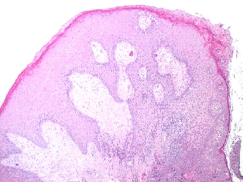
Microscopic image of the gingival mucosa showing its overall hypertrophy. On the surface, there is a thickened gingival epithelium, with deep epithelial ridges, acanthosis, and hyperkeratosis in the superficial layers. HE staining, ×40

Microscopic image of the gingival mucosa showing its overall hypertrophy. On the surface, there is a thickened gingival epithelium, with deep epithelial ridges, acanthosis, and hyperkeratosis in the superficial layers. HE staining, ×40