Figure 6.

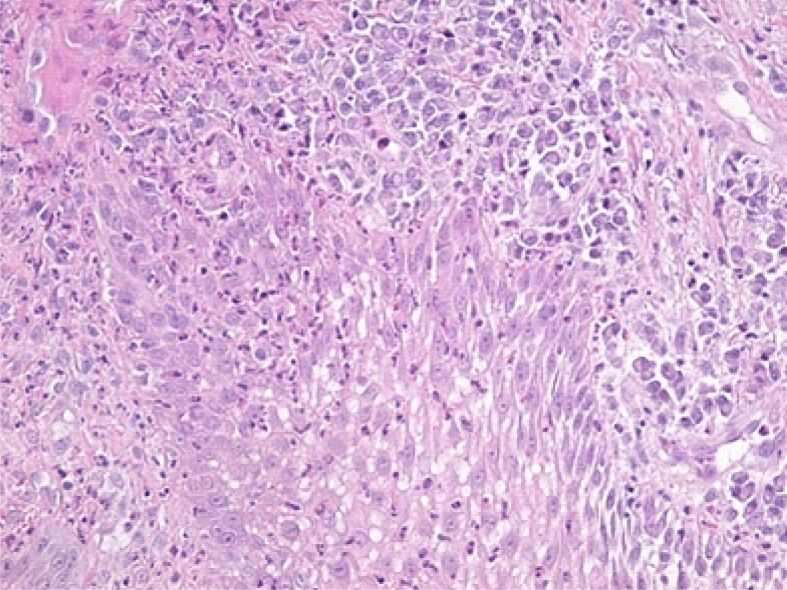
Microscopic image from the deep area of the gingival epithelium showing the presence of an intraepithelial edema and infiltration with lymphocytes and neutrophilic granulocytes. HE staining, ×200

Microscopic image from the deep area of the gingival epithelium showing the presence of an intraepithelial edema and infiltration with lymphocytes and neutrophilic granulocytes. HE staining, ×200