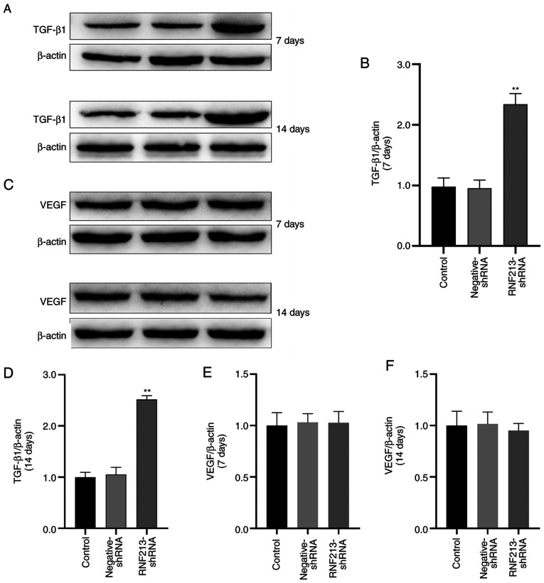
Figure 7.
Detection of TGF-β1 and VEGF protein levels in rBMSCs following RNF213 silencing. (A) Representative western blotting images of TGF-β1 expression 7 and 14 days after transfection. (B) TGF-β1 expression 7 days after transfection was quantified. (C) Representative western blotting images of VEGF expression 7 and 14 days after transfection. (D) TGF-β1 expression 14 days after transfection was quantified. VEGF expression (E) 7 and (F) 14 days after transfection was quantified. **P<0.001 vs. Control. TGF-β1, transforming growth factor β1; VEGF, vascular endothelial growth factor; rBMSCs, rat bone marrow mesenchymal stem cells; RNF213, ring finger protein 213; shRNA, short hairpin RNA.

