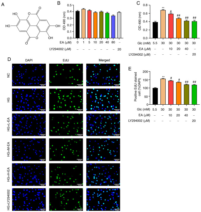
Figure 1.
EA inhibits the proliferation of MCs induced by HG. (A) Chemical structure of EA. (B) Cytotoxicity of EA (1-80 µM) and LY294002 (20 µM) was detected via MTT assay in MCs treated with 5.5 mM glucose. (C) Effect of EA (10, 20 and 40 µM) and LY294002 (20 µM) on MC viability was detected using MTT assay. Effect of EA (10, 20 and 40 µM) and LY294002 (20 µM) on MC proliferation was (D) detected by EdU assay and (E) quantified. Scale bar, 40 µm. **P<0.01 vs. NC group; #P<0.05 and ##P<0.01 vs. HG group. normal control (NC) group refers to 5.5 mM glucose treated cells. EA, ellagic acid; MCs, mesangial cells; OD, optical density; HG, high glucose; H-EA, high concentration EA intervention group; M-EA, medium concentration EA intervention group; L-EA, low concentration EA intervention group; NC, normal control; Glc, glucose; EdU, 5-ethynyl-2'-deoxyuridine.

