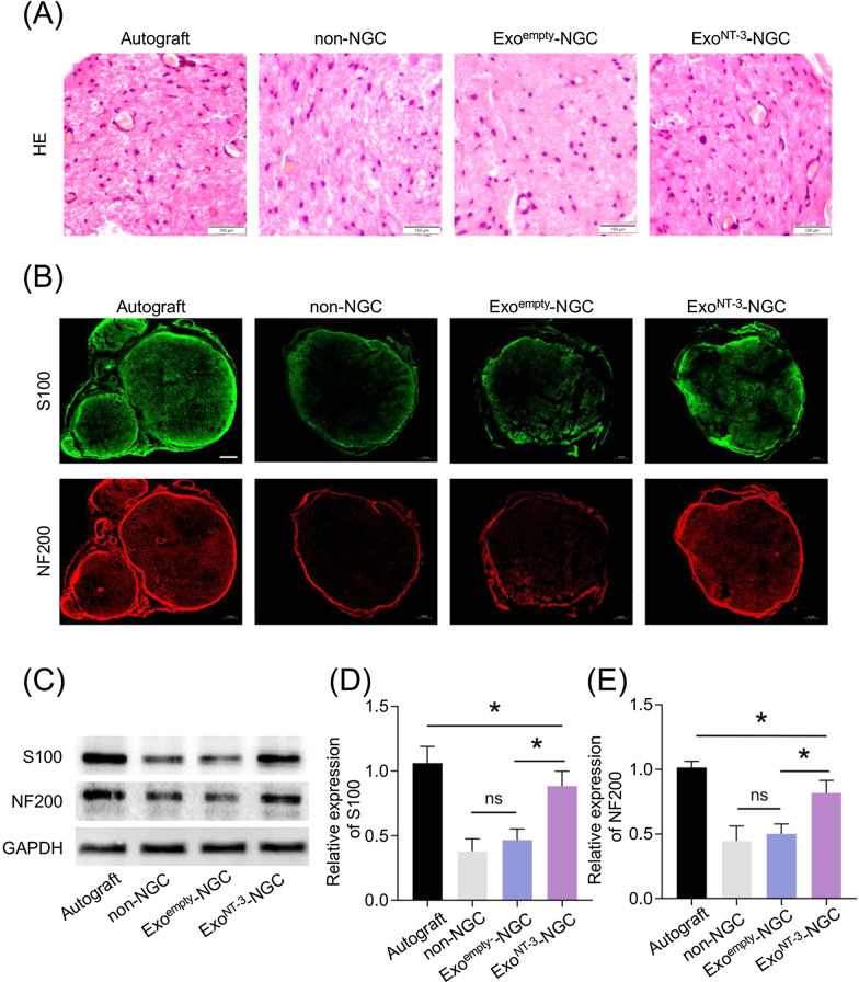
Fig. 4.
Evaluation of the regenerated nerves morphology and immunofluorescence analysis. a Cross section from the distal segments of regenerated nerves were stained by HE stain. The blue arrow indicates vessels. b Cross section from the distal segments of regenerated nerves were immunostained 8 weeks after the implantation. S100 indicated the regenerated SCs and NF200 indicated the regenerated axons. Scale bar = 100 μm. c Representative western blotting of S100 and NF200 protein expressing in regenerated nerve tissues. d Statistical analysis of S100 protein expression in regenerated nerve tissues. e Statistical analysis of NF200 protein expression in regenerated nerve tissues. Data are presented as mean ± SEM, n = 6 rats per group, *P < 0.05 by ANOVA with post hoc Bonferroni correction

