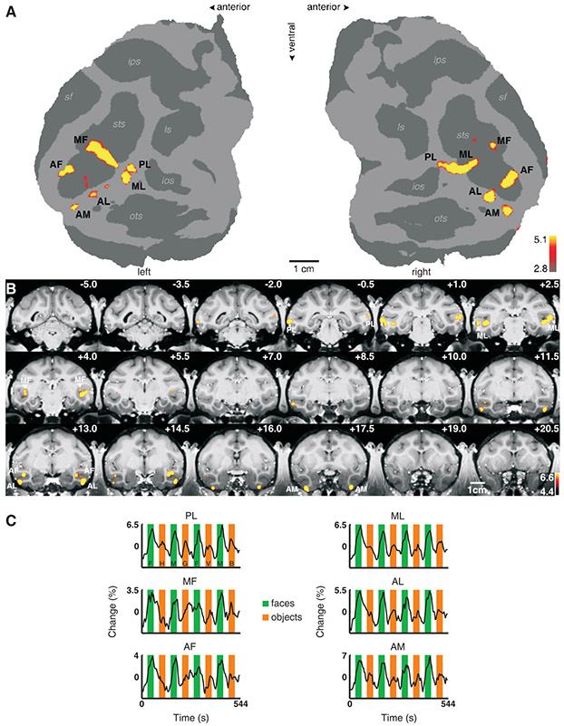
Fig. 1.
Face-selective patches in monkey M1. (A) The flattened cortical surfaces (“flatmaps”) from both hemispheres show regions significantly more activated by faces than by other objects. Computational flattening involves distorting the spatial arrangement of the data, and under-estimates the size of the sulci (shown in dark grey). Anatomical labels: sts: superior temporal sulcus, sf: Sylvan fissure, ips: intraparietal sulcus, ls: lunate sulcus, ios: inferior occipital sulcus, ots: occipitotemporal sulcus. (B) The same contrast overlaid on high resolution coronal slices from monkey M1. The anterior-posterior position of each slice in mm relative to the interaural line is given in the top right corner; the left hemisphere is shown on the left. The face patches are labeled as in (A). (C) Mean time courses extracted from the six face patches of the right hemisphere. Three different visual stimulation conditions were presented: faces (green epochs, F: human faces, M: monkey faces), objects (orange epochs, H: hands, G: gadgets, V: vegetables and fruits, B: monkey bodies), and scrambled versions of the same images (white epochs).

