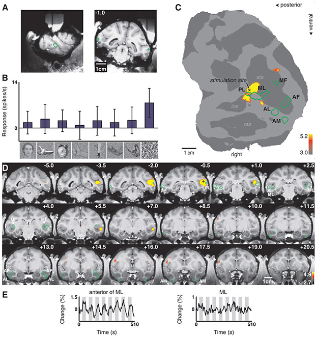
Fig. 4.
Brain regions activated by microstimulation outside the lateral middle face patch (ML) in monkey M1. Same conventions as Fig. 2. (A) Electrode position just posterior to ML. (B) Example of neuronal selectivity. (C) The contrast microstimulation versus no microstimulation revealed microstimulation-induced activity around the stimulation site as well as in a distinct patch anterior to ML. (D) The same contrast overlaid on coronal slices. Note how the activation spared most of PL and the other face patches. (E) Time courses from the patch just anterior to ML and from ML.

