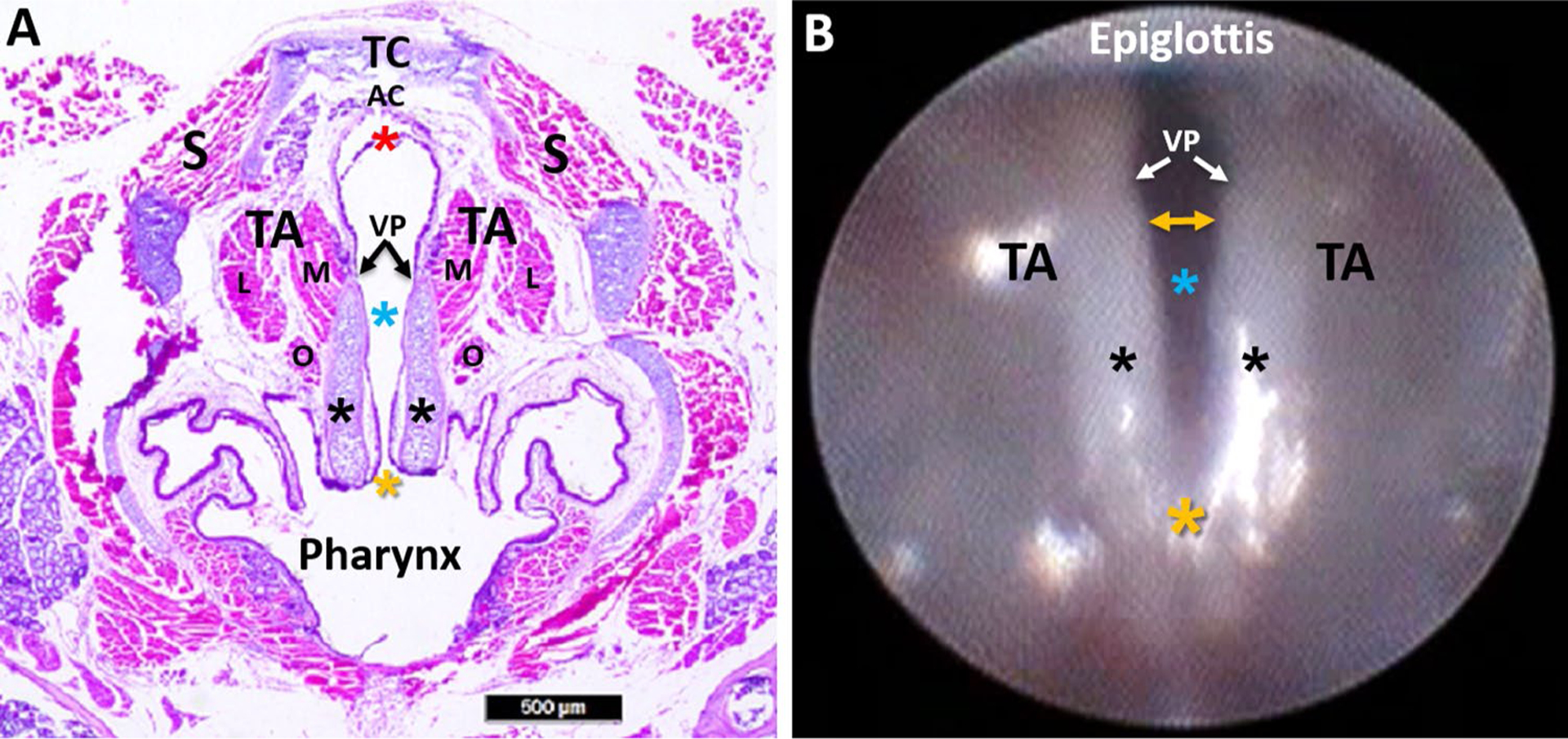
Fig. 10.

Murine laryngeal framework. a Hematoxylin and eosin (H&E) stained transverse Sect. (10 μm) of a mouse larynx at the level of the VFs, with labeled structures. b Endoscopic image of the murine VFs, with corresponding labeled structures from image a. In contrast to the human larynx, mice have proportionately larger arytenoid cartilages (black asterisks) and a proportionately smaller mucosal region extending beyond the vocal processes (VP) to the ventral commissure (red asterisk). In the mouse, the ventral commissure (which is obscured during laryngoscopy) is framed by a U-shaped alar cartilage (AC) that does not exist in humans. During spontaneous breathing in the mouse, the most dorsal portion of the arytenoids (yellow asterisk; dorsal commissure) remains relatively fixed near midline, serving as a pivot point for VF abduction and adduction. As a result, VF movement in the mouse is more readily apparent at the ventral (yellow bidirectional arrow in image b rather than the dorsal (posterior) region as in humans. TC thyroid cartilage, AC alar cartilage, VP vocal process, TA thyroarytenoid muscle (M medial belly, L lateral belly, O oblique belly), S strap muscles; blue asterisk: glottis; black asterisk: arytenoid cartilage; yellow asterisk: dorsal commissure; red asterisk: ventral commissure. Scale bar 500 μm
