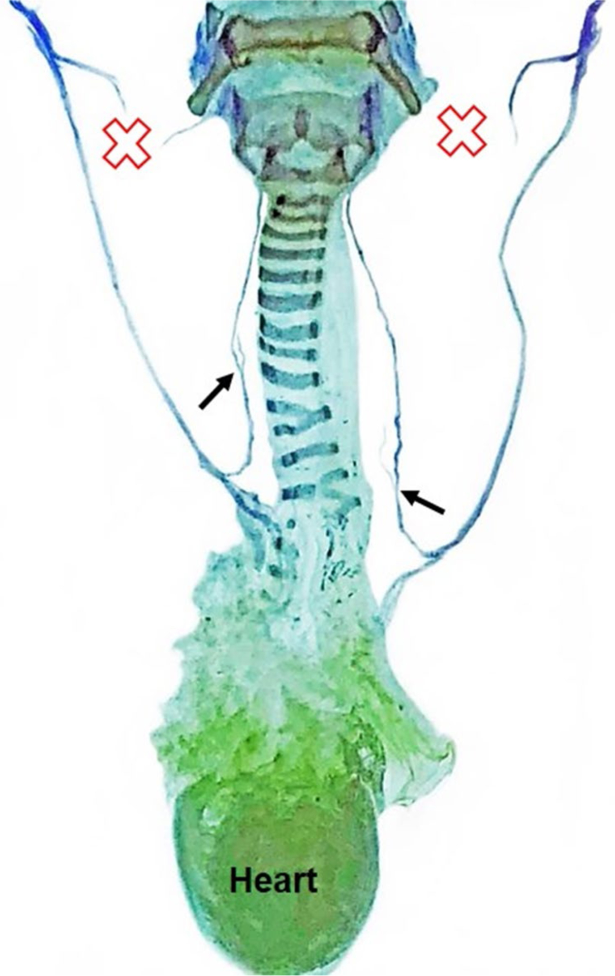
Fig. 8.

Sihler staining for laryngeal nerve mapping. A representative Sihler stained sample from a mouse in the bilateral SLN transection group demonstrates that the murine laryngeal framework and laryngeal nerve branching pattern are remarkably similar to humans. Red X indicates the location of SLN transection. Black arrows show the origin of nerve branches from the RLN trunk bilaterally. CN X Cranial nerve 10 (i.e., vagus nerve), RLN recurrent laryngeal nerve, SLN superior laryngeal nerve
