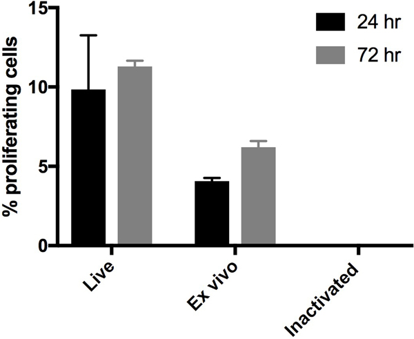
Figure 1.
4T1 tumor cells were injected into the mammary fat pad of Balb/c mice. When tumors reached ~10mm in diameter, the mice were euthanized and tumors were removed, and processed into a single cell suspension. Some of the tumor cells were inactivated using RF+UV (“Inactivated”). Some of the tumor cells were placed directly in culture (“Ex vivo”). The “Ex vivo” and “Inactivated cells,” along with the original 4T1 tumor cell line (“Live”) were incubated at 37C for 24 or 72 hours. Proliferation was measured using EdU staining, where the EdU was added during the last 24 hours of culture.

