
Figure 7.
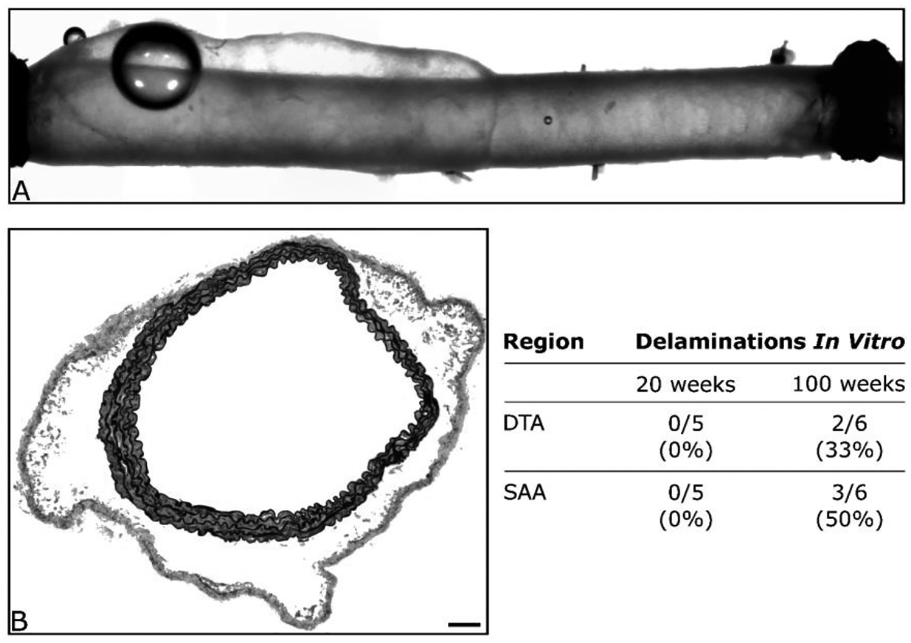
Shown are an illustrative (panel A) video-capture during standard biaxial testing of a DTA that was excised from a 100-week old mouse and (panel B) histological image of a SAA following mechanical testing. Both images reveal a propensity to develop an intramural delamination without frank rupture. Such delaminations consistently occurred at the medial-adventitial border and appeared to initiate at or near branches. The scale bar represents 100 μm in panel B.