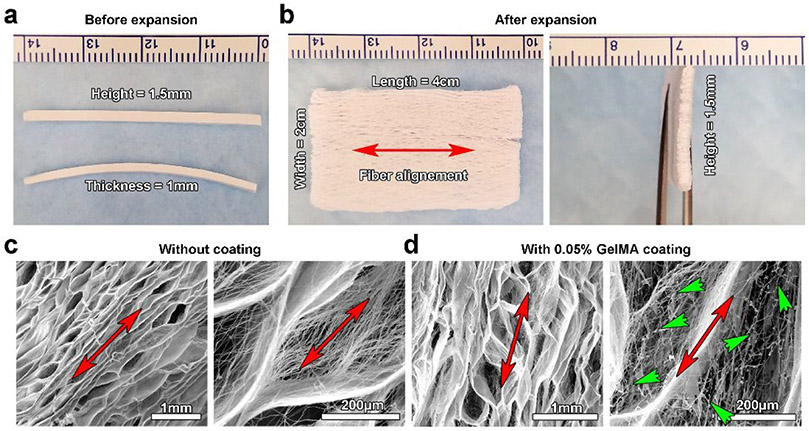
Figure 2.
3D expanded nanofiber scaffold fabrication and characterization. (a) Photograph showing PCL nanofiber stripes (4 cm × 1.5 mm × 1 mm) before expansion. (b) Photograph showing a 3D expanded PCL nanofiber scaffold (4 cm × 1.5 mm × 2 cm). (c) SEM images showing 3D expanded PCL nanofiber scaffolds without 0.05% GelMA coating, indicating the porous and layered structure. (d) SEM images showing 3D expanded PCL nanofiber scaffolds with 0.05% GelMA coating, indicating the formation of hybrid nanofiber scaffolds with a porous and layered structure. Red arrows indicate the direction of fiber alignment. Green arrow heads indicate the formation of GelMA nanofibers.

