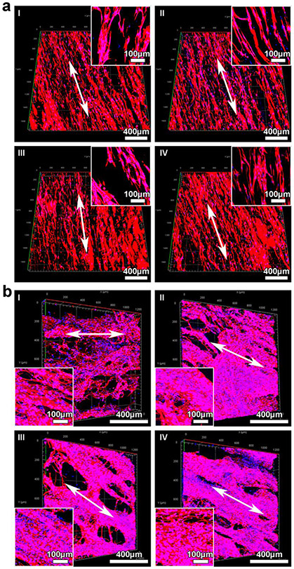
Figure 4.
Cell culture on 3D expanded nanofiber scaffolds (2 cm × 2 cm ×1.5 mm). (a) Conformal microscopy images showing BMSCs in different regions of square-shaped nanofiber scaffolds. The depth imaged was 125 μm. Inset: highly magnified images of BMSCs seeded on nanofiber scaffolds. Cells were stained with Alexa Fluor™ 546 Phalloidin in red, and cell nuclei were stained with DAPI in blue. (b) Conformal microscopy images showing hNSCs in different regions of square-shaped nanofiber scaffolds after proliferation for 7 days and neuronal differentiation for another 14 days. Inset: The formation of 3D neuronal networks on the scaffolds after neuronal differentiation for 14 days. Cells were stained with Tuj1 for neurons in red, and cell nuclei were stained with DAPI in blue.

