Figure 5.
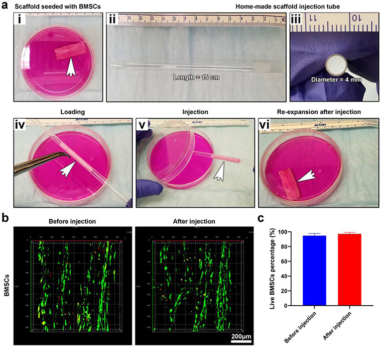
Demonstration of minimally invasive delivery of 3D tissue constructs and the effect of delivery on the cell viability. (a (i)) Photograph showing a 3D tissue construct consisting of 0.05% GelMA-coated, 3D expanded nanofiber scaffolds seeded with BMSCs in a petri dish. (a (ii, iii)) Photographs showing glass tubes for injection of 3D tissue constructs. (a (iv, v)) Photographs showing the 3D tissue construct was loaded into the glass tube and then injected into the petri dish containing culture medium using an air source. (a (vi)) Photograph showing the injected 3D tissue construct returned to its original shape. (b) LIVE/DEAD staining of BMSCs seeded on 0.05% GelMA-coated, 3D expanded nanofiber scaffolds before and after injection. (c) Quantification of live BMSCs before and after injection.

