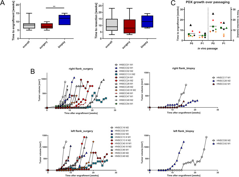
Fig. 3.
PDX formation, success rate, and growth kinetic in NSG mice. A Boxplots depicting the time from implantation to engraftment (appearance of palpable tumor in the flanks) and the time from the engraftment to resection when tumors reached maximum sizes [1500 mm3]. Whiskers show the minimum and maximum. **p < 0.01, unpaired t-test (two tailed). B Growth curves of the individual PDX: Each line represents a PDX grown in P0 in one mouse. M – mouse. C Growth acceleration after passaging: Scatterplot depicting the time from implantation to engraftment and resection for the initial implantation (= P0) and after cryopreservation and replantation into NSG mice (= P1). Each dot is representative for an individual HNSCC case using the same symbol and color for P0 and P1

