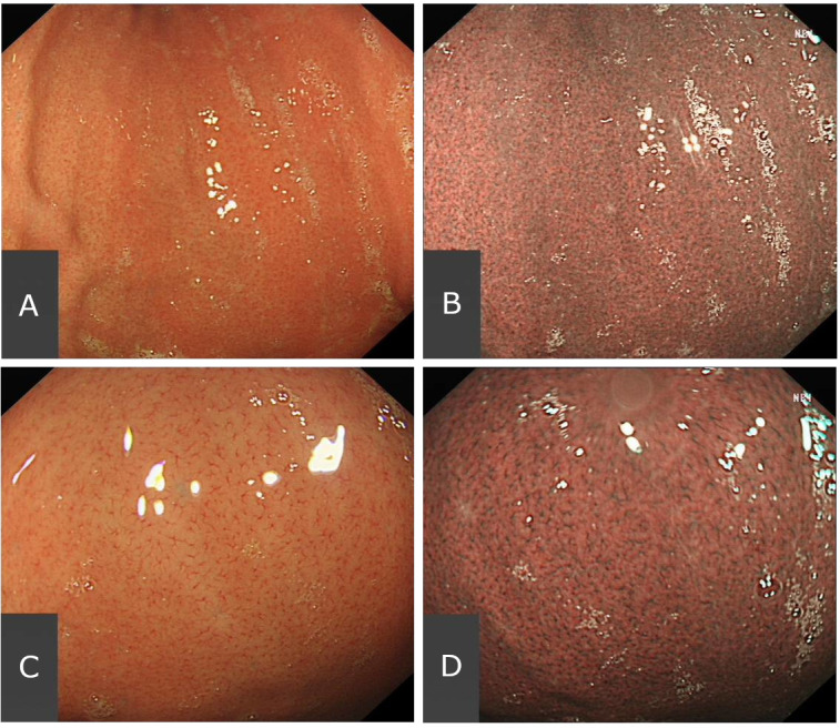
Figure 1.

The endoscopic appearance of the RAC in the distal stomach, in a Helicobacter pylori naïve patient. Examined at medium distance without magnification under white-light endoscopy (WLE) (A) and narrow-band imaging (B), and on closer inspection without magnification under WLE (C) and NBI (D). NBI, narrow-band imaging.
