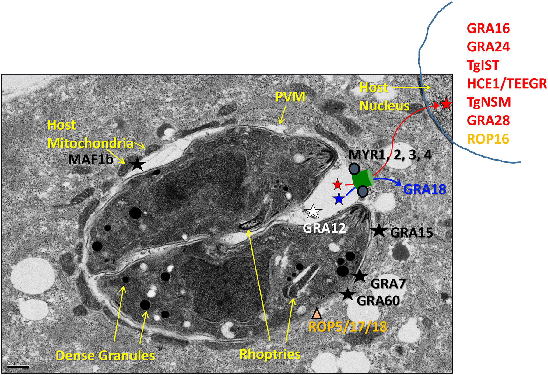
Figure 1.

Toxoplasma dense granule effector proteins and their functional locations. After infection, Toxoplasma dense granules secrete a large number of proteins into the parasitophorous vacuole, some of which give the PV unique structure, others of which function in metabolism, and yet others of which are termed effector proteins and localize to various cellular compartments to interact with the host cell. Effectors that transit the PV and then exit via a MYR-dependent mechanism (conceptualized by a green channel but with an as-of-yet unknown structure and mechanism of operation) are then located in the host cytosol (blue) and the host nucleus (red). Effector proteins that contain a transmembrane domain are inserted into the PVM via a MYR-independent mechanism which remains unknown, and are indicated by black. One effector, GRA12, is indicated by white and remains within the PV, associating with the membranous network therein, where it nevertheless has an important impact on host cell functions. The rhoptry proteins ROP5/17/18 that sit on the PVM and interact with GRA effectors are also shown in orange; ROP17 also has a role in the export of PVM-transiting, MYR-dependent GRA effectors.
