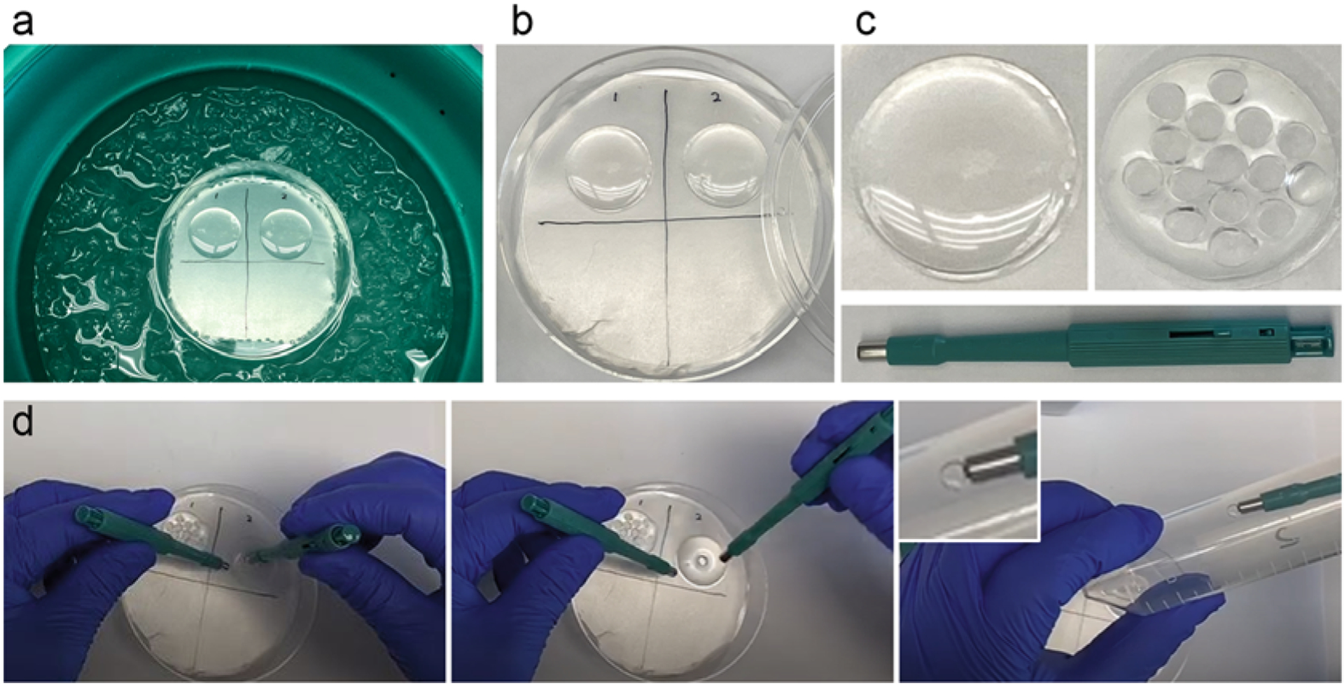
Fig. 3.

Polymerization and punching. (a) A 100-mm petri dish with its bottom layered by Parafilm on the top of an ice–water bath. Two coverslips with cells facing up are covered with polymerization mixture. Samples are kept on ice during the first 20 min of polymerization, followed by incubation at RT for another 1–2 h (b). (c) A coverslip containing cells and polymerized gel before (left) and after (right) samples have been excised using a 4-mm biopsy puncher (green, below). Multiple individual samples can be excised from one gel. (d) Punching and transferring of a punch to a 50 mL conical tube
