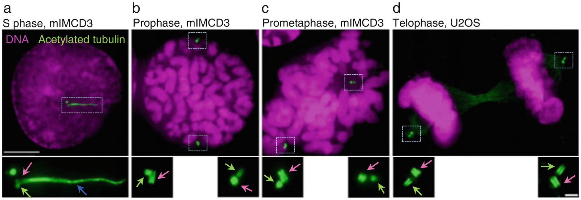
Fig. 6.

Examples of expanded centrioles and cilia using herein described method. Cells were grown on coverslips, expanded ~4.2×, and immunolabeled using an antibody recognizing acetylated tubulin. DNA is labeled with DAPI. (a) An interphase cell with two mother centrioles (pink arrow) associated with procentrioles (green arrow). One mother centriole is ciliated (blue arrow is pointing to the ciliary axoneme). (b–d) Examples of cells in various stages of mitosis. Each mother centriole (pink arrows) is associated with one procentriole (green arrows). Centrioles are in various orientations. Scale bars: 20 and 2 μm for the inserts
