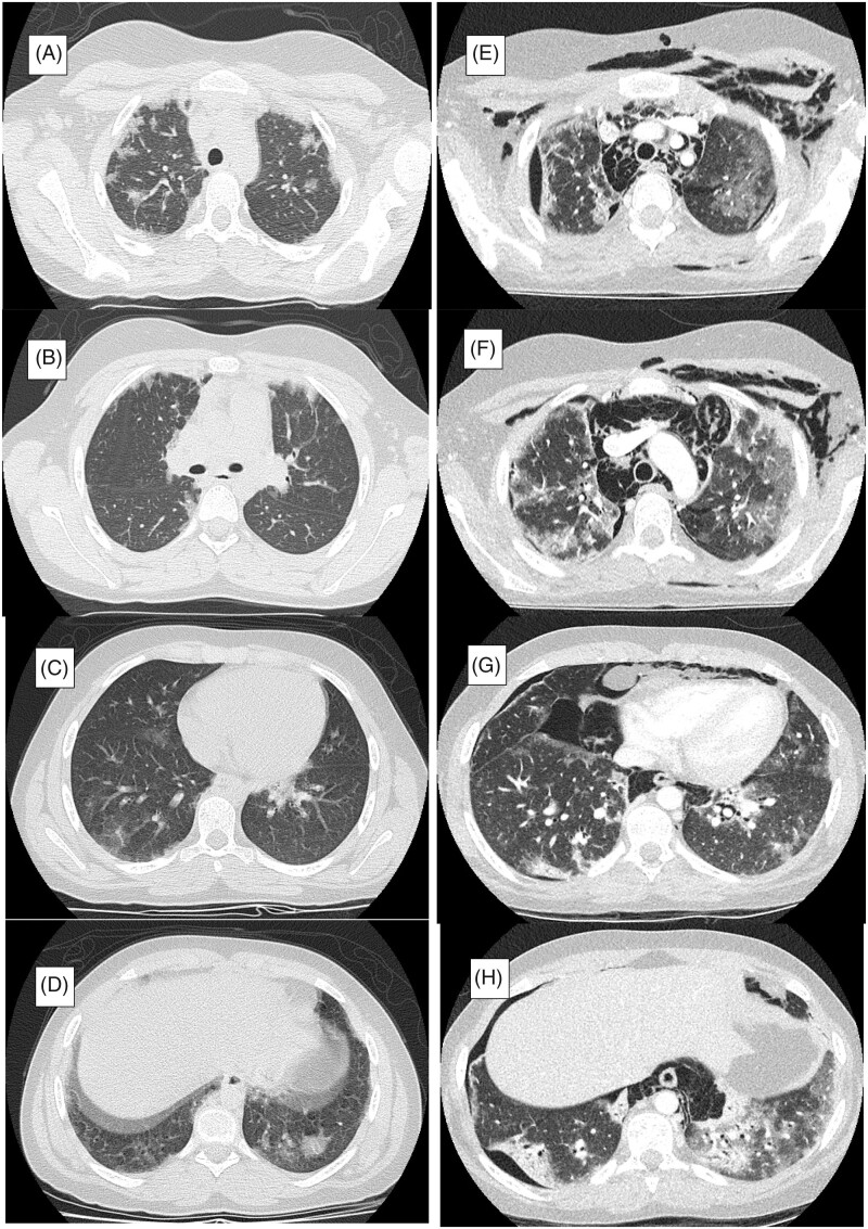
Figure 3.
Time course of chest CT images. (A–D) Multifocal, bilateral and peripheral predominant ground glass opacities were found on chest computed tomography (CT) at the first admission in the previous hospital. (E–H) Chest CT at PICU admission in our hospital. Severe RP-ILD (peripleural ground-glass opacity and patchy distribution of areas of consolidation accompanied by traction bronchiectasis) further complicated with pneumomediastinum, pneumothorax and cervical subcutaneous emphysema. PICU: Paediatric Intensive Care unit; RP-ILD: rapidly progressive interstitial lung disease.

