Figure 2.

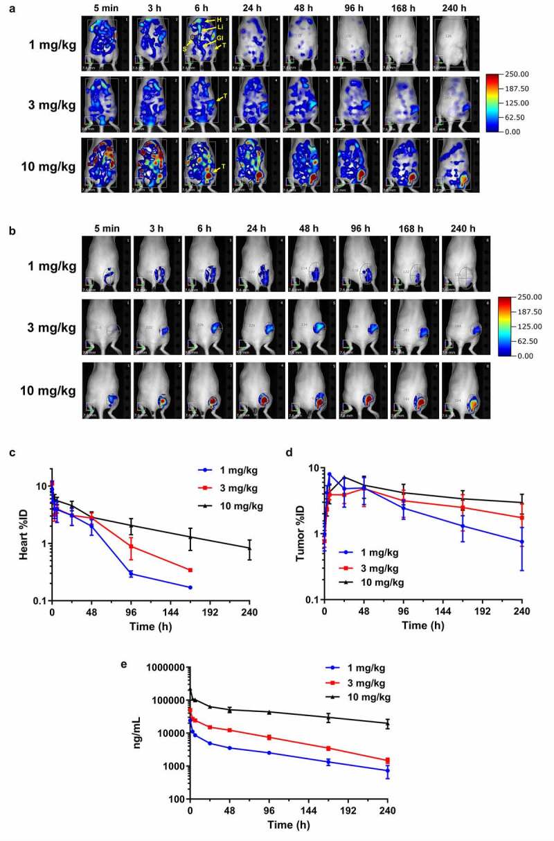
In vivo biodistribution and tumor targeting by FMT imaging. (a) noninvasive longitudinal FMT imaging of whole body at 5 min, 3, 6, 24, 48, 96, 168, and 240 h after injection of anti-IL13Rα2-Ab-AF680 in A375 xenograft-bearing mice at 1, 3, or 10 mg/kg doses. Shown are time course images of one representative mouse per group. T – tumor; H – heart; Li – liver; S – spleen; Gi – gastrointestinal tract. (b) Tumor regions of interest (ROIs) demonstrating targeting profile and increased fluorescence signal with higher doses. (c-d) In vivo ROI quantitation of heart (c) and tumor (d) uptake (%ID) in various dose groups. (e) Plasma pharmacokinetic profile of anti-IL13Rα2-Ab at different doses as evaluated by ligand binding assay. N = 3– 6 mice per group for each time point; data are Mean ± SEM
