
Figure 7: Effect of cycloheximide on VE-Cad (media and lysate) and ZO-1 (lysate) at 18 hr.
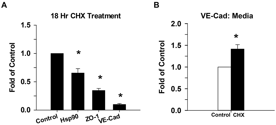
P-UAEC grown to near confluence were treated with cycloheximide (CHX), a protein synthesis inhibitor, for 18 hr. The expression of VE-Cad and ZO-1 were then measured in cell lysates by western blot, and normalized to Hsp90 (Panel A). Shed VE-Cad expression was also measured in the media (Panel B) and was normalized to nonspecific staining of BSA. Data is consistent with findings for changes in both VE-Cad vs ZO-1 in Figs 3 and 4. Values shown are mean ±SEM of 4–5 independent experiments and expressed as fold of control. *P<0.05 compared with control; +P<0.05 compared with same agonist treatment.