Figure 2.
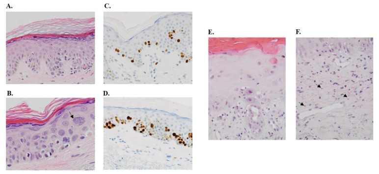
Histopathological changes in the skin. (A) This shows a skin biopsy of a patient after treatment with CPC634 with epidermis demonstrating hypergranulosis, influx of lymphocytes, basal vacuolisation of keratinocytes and scattered apoptotic cells along the dermo-epidermal junction (20× magnification). (B) This section is a 40× magnification of the same patient showing keratinocyte micronucleation (“grape cells”, arrow), also notice the accompanying apoptosis. Immunohistochemical stain for Ki67 at baseline (C) and after CPC634 treatment (D) (20× magnification). (E) This skin biopsy section of a patient in the Napoly study with skin toxicity shows an epidermis demonstrating vacuolar interface dermatitis with basal vacuolisation, influx of lymphocytes, scattered high apoptosis and hyper-and parakeratosis (20× magnification). Note the keratinocyte atypia with polymorphic nuclei and prominent nucleoli. (F) This section of the same patient shows ectatic capillaries and postcapillary venules of the superfical vascular plexus, a minor lymphohistiocytic perivascular infiltrate and extensive erytrocyte extravasation (arrow) (20× magnification).

