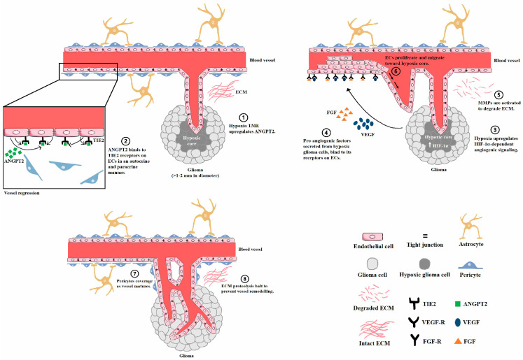
Figure 1.
Schematic representation of glioma angiogenesis. As tumor growth within the brain parenchyma goes beyond 1–2 mm in diameter, its metabolic demands cannot be met entirely via diffusion. (1) Hypoxia then occurs to regulate angiogenic signals. (2) The activation of autocrine and paracrine ANGPT2/TIE2 signaling subsequently disrupts endothelial–mural cell interactions for vessel regression. (3,4) HIF-1α upregulation induces the transcriptional activation of proangiogenic molecules that initiate EC proliferation and migration. (5) Simultaneously, increased ANGPT2 activates MMP-2 to degrade the ECM. (6) Proteolysis of the vessel’s basement membrane facilitates the migration and proliferation of ECs toward the hypoxic tumor core. Lastly, the blood vessel wall matures, as (7) pericytes are recruited along the ECs to stabilize the blood vessel. (8) Endogenous protease inhibitors and antiangiogenic factors locally halt ECM proteolysis to hinder further vessel remodeling.

