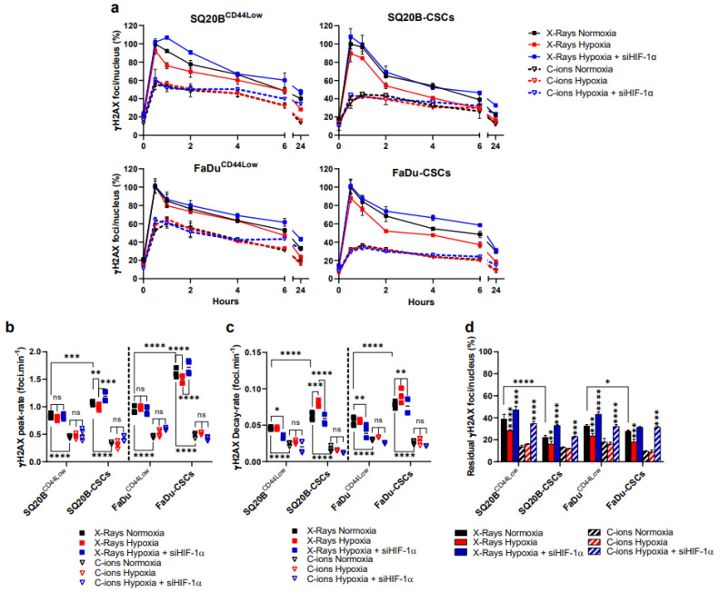
Figure 3.
Silencing HIF-1α under hypoxia increases the residual detected DSBs in response to both irradiations. (a) Kinetics of γH2AX foci after X-rays and C-ions, ± siHIF-1α. The percentages of foci per nucleus were calculated for each condition considering the X-ray normoxic peak as the reference (means ± SD). (b,c) represent respectively the peak-rate from basal levels and decay-rate from peak to 6 h, as determined from the data shown in (a) (two-way ANOVA test, * p < 0.05, ** p < 0.01, *** p < 0.001, **** p < 0.0001, ns: non-significant). (d) Residual foci were quantified at 24 h and each condition was statically compared to their respective normoxic condition (two-way ANOVA test) (n = 3).

