Figure 1.
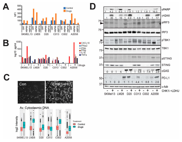
CHK1i+LDHU increases markers of ICD. (A) Human melanoma tumourspheres were treated with or without 0.2 μM SRA737 + 0.1 mM HU and harvested at 48 h for analysis of the cell surface expression of calreticulin (CRT), HSP70 and HSP90 on live cells. These data are representative of two independent experiments. (B) The indicated human melanoma tumoursphere lines were treated with or without 0.1 μM GDC-0575 + 0.1 mM HU for 48 h and the tissue culture supernatants were harvested and the cytokine levels measured. These data are representative of two independent experiments. (C) Immunostaining for cytoplasmic DNA in A2058 melanoma cells without and with treatment with 0.1 mM GDC-0575+0.1 mM HU (CHK1i+HU) for 48 h. Bar indicates 10 mm. The indicated melanoma cell lines were grown on plastic and treated as in B then fixed and stained for cytoplasmic DNA, and the level of staining was quantitated using high content imaging. The data are the mean and 95% CI from triplicate samples. (D) The indicated tumoursphere cultures were treated as in B and harvested at 24 h for immunoblotting of the indicated markers. The change in band intensity relative to the no-drug control for each cell line are shown. Where no changes are observed no quantitation is shown.

