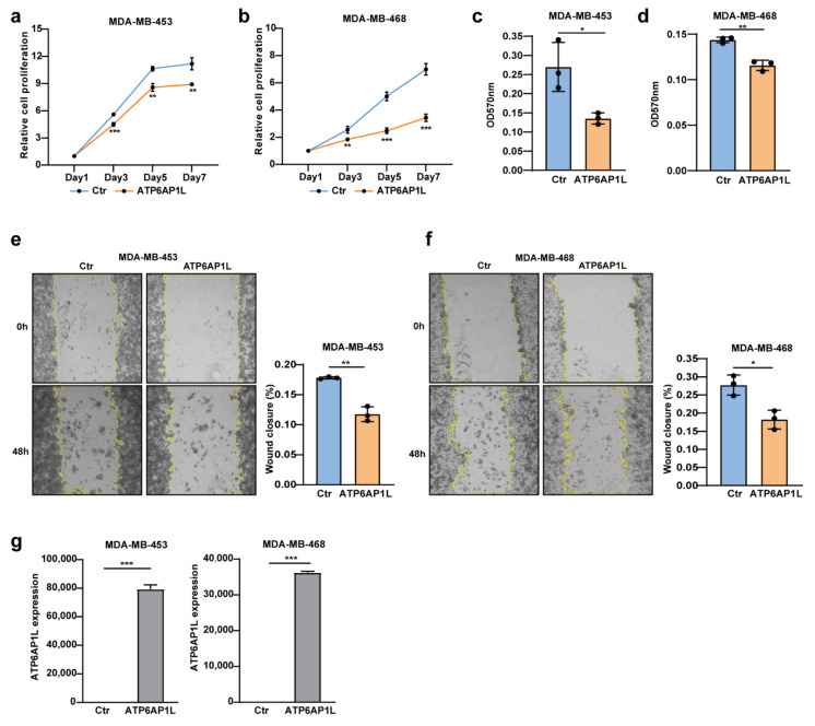
Figure 5.
ATP6AP1L overexpression suppresses TNBC cell proliferation and migration. (a,b) Cell proliferation assay of two TNBC cell lines, MDA-MB-453 (a) and MDA-MB-468 (b), which were overexpressed with the ATP6AP1L gene by lentiviral infection. Empty vector packaged virus was used as the control. Cells numbers were measured at the indicated time points with the CCK-8 assay and represented as absorbance at 450 nm. Values are the means ± SD; ** p < 0.01, *** p < 0.001, two-tailed Student’s t-test. (c,d) Colony-forming assay for MDA-MB-453 (c) and MDA-MB-468 (d) cells that were overexpressed with the ATP6AP1L gene by lentiviral infection. Colonies were stained with crystal violet and quantified by reading absorbance at 570nm. Values are the means ± SD; * p < 0.05, ** p < 0.01, two-tailed Student’s t-test. (e,f) The wound-healing assay images for MDA-MB-453 (e) and MDA-MB-468 (f), which were overexpressed with the ATP6AP1L gene by lentiviral infection. The histogram on the right depicts the quantification of the wound closure percentage fraction. Values are the means ± SD; * p < 0.05, ** p < 0.01, two-tailed Student’s t-test. (g) ATP6AP1L overexpression in MDA-MB-453 and MDA-MB-468 cells was analyzed by qPCR. Values are the means ± SD; *** p < 0.001, two-tailed Student’s t-test.

