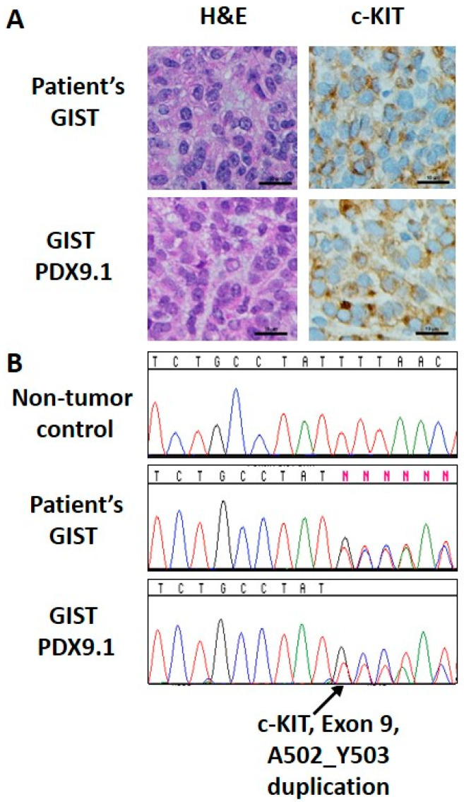
Figure 5.
Immunohistochemical and mutational analysis of a primary GIST and matched PDX model. (A) H&E and c-KIT (CD117) staining for primary GIST (top) and matched GIST PDX9.1 (bottom). (B) Sanger sequencing analysis identified the A502_Y503 duplication in exon 9 of KIT in both primary GIST (middle) and matched GIST PDX9.1 (bottom), but not in a non-tumor specimen (top). Scale bar: 10 μm.

