. 2021 Aug 6;44(3):e20200332. doi: 10.1590/1678-4685-GMB-2020-0332

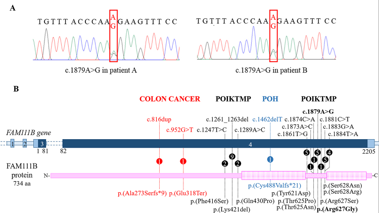
Figure 2 -. The mutational repertoire of FAM111B. A) Electropherograms showing the c.1879A>G FAM111B missense variant (framed by the red rectangle) detected in the two unrelated Italian patients. B) Picture summarizing the FAM111B mutational repertoire. The dark blue bar indicates the coding regions of the gene while the non-coding regions are in light blue. The pink bar indicates the protein: the punctate boxes correspond to the protease domain, spanning from amino acid residues 471 to 664 and interrupted by a linker region (dotted bar). Each change is described at DNA (top) and protein level (below). Numbers within the circles specify the number of patients carrying the variant. Pathogenic variants associated with POIKTMP, POH, and colon cancer are written in black, blue and red, respectively.

Figure 2 -