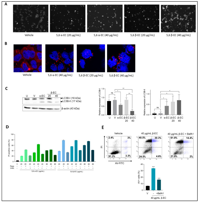
Figure 5.
5,6-ECs induce an autophagy-mediated cell death. (A) U266 cells were treated with vehicle or 5,6 α/β-EC (20, 40 µg/mL) for 20 h. Cells were then stained with an autophagosome specific fluorescent probe and analyzed with an Olympus BX53 fluorescent microscope (× 40, magnification) (B) p62 expression was analyzed by IF in EtOH- and 5,6-ECs-treated cells. We used a primary Ab against p62, and a goat Alexa Fluor 488-conjugated anti-rabbit IgG as secondary Ab. Slides were counterstained with DAPI and analyzed with a confocal microscope (Fluoview FV100, Olympus, CA, USA) (× 180, magnification). (C) U266 cells were untreated (U), vehicle-treated (E) or with 5,6 α-EC (40 µg/mL) or 5,6 β-EC (20 or 40 µg/mL). Within 24 h, whole-cell proteins were extracted, separated by SDS-PAGE, and transferred onto membranes then incubated with anti-LC3B or anti-β-actin (as a control) antibodies. Protein levels were estimated by densitometry and collected data from three independent experiments were presented in histograms (means ± SD). (D). The modification of autophagic flux was evaluated by flow cytometry. U266 cells were seeded into 24-well plates for 24 h. The cells were treated with 5,6 α-EC or 5,6 β-EC alone or in combination with 5 µM rapamycin or 10 µM 3-MA for 24 h. The cells were stained with PI and PI positive cells were recorded. At least 104 events were gated. The experiment has been done once with triplicate samples. (E) U266 cells were pre-treated with BafA1 (50 nM) for 4 h and then treated with 5,6 β-EC (40 μg/mL). The cells were stained with annexin V/IP as described before and sorted. Cytometry profiles were presented together with the percentage of cells within each quadrant. The percentage of annexin V+ cells in each culture condition is presented in the histogram. * p <0.05; ** p < 0.01; *** p < 0.001; ns, not significant with the t-test.

