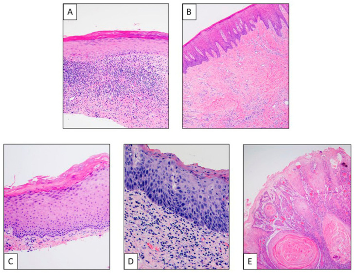
Figure 2.
Different histological entities of oral lesions. (A) Lichen planus: Histologically characterized by degeneration of the basal cell line of the epithelium oral mucosa, with presence of diverse degrees of orthokeratosis and parakeratosis besides thickness of spinous layer and a characteristic band-like lymphocytic infiltration at the basal level and superficial submucosa. (B) Oral submucous fibrosis: This is a chronic progressive scarring lesion, morphologically showing evident submucosal changes such as fibrosis, diffuse chronic inflammatory infiltrate, atrophy of minor salivary glands, skeletal muscle atrophy, band-like infiltrate, edema and congestion, and vesicle formation. At mucosa level, we found changes such as atrophic changes, pigment incontinence, ulceration with granulation tissue, hyperplastic changes, dysplasia or malignant transformation. (C) Low-grade dysplasia. (D) High-grade dysplasia/in situ carcinoma: Dysplasia is a premalignant condition that refers to abnormal epithelial growth characterized by architectural and cytologic atypia, including dyskeratosis, basal cell hyperplasia and anaplasia, keratin pearls, etc. Low-grade refers to architectural and cytologic atypia affect from 1/3 to 2/3 of epithelium; high-grade/in situ carcinoma represents full thickness cytological or architectural atypia, without invasion of the neoplastic keratinocytes through the basement membrane. (E) Infiltrating squamous cell carcinoma: Malignant neoplasm characterized by severe dysplasia of the surface epithelium with invasion of the stromal–epithelial interface.

