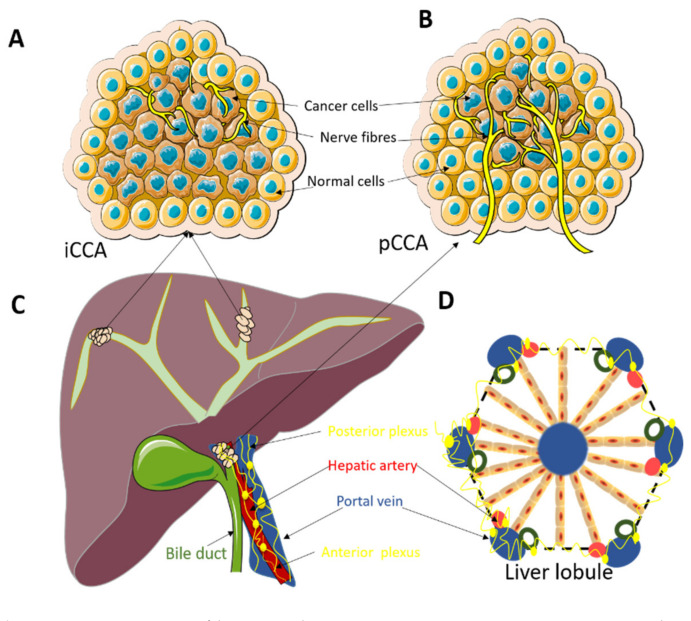
Figure 1.
(A): Nerve fibers in the tumor microenvironment in intrahepatic cholangiocarcinoma (iCCA). The small nerve fibers are mainly distributed at the edge of the tumor. (B): Nerve fibers in the tumor microenvironment in perihilar cholangiocarcinoma (pCCA). The small nerve fibers are growing in between the tumor glands and not just at the edge of the tumor as in iCCA. (C): Localization of iCCA and pCCA tumors. ICCA is located centrally in the liver and usually presents with a big tumor mass. PCCA are located at the liver hilum and usually present as smaller tumors. (D): Architecture of the normal liver lobule. The normal innervation of the liver lobule can possibly explain the observation of the presence of the small nerve fibers at the edge of the tumor in iCCA and the pattern of mixed distribution of small nerve fibers in pCCA.

