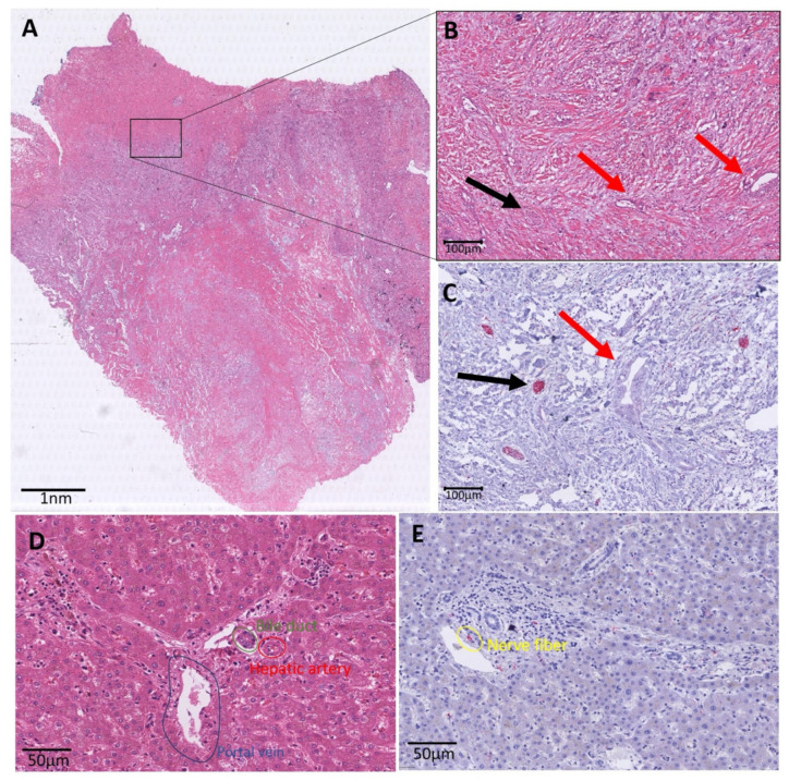
Figure 4.
Histology overview of iCCA. (A): Whole slide H&E image of an iCCA. At the edge of the slide, normal liver parenchyma is displayed. More centrally, a lesion is shown with a rim of vital tumor cells, and centrally, a pale area corresponding with necrosis. (B): Zoomed in image of the black marked box in A. This H&E image shows small tumor glands in abundant stroma marked with red arrows. The black arrow points to a nerve fiber, which is not easily visible on this HE staining. (C): Black box area in the immunohistochemical PGP9.5 staining. This staining makes it easier to recognize the small nerve fibers (red in the PGP9.5 immunohistochemistry and marked with a black arrow). The red arrow is pointed at the tumor. (D): Zoomed in image of a portal tract. The portal tract illustrates the bile duct (marked in green), the hepatic artery (marked in red) and the portal vein (marked in blue). (E): Zoomed in image of a portal tract with PGP9.5 staining. In this zoomed in image, the small nerve fiber is nicely illustrated in red (positive immunohistochemical staining) and marked in yellow.

