
Fig. 8.
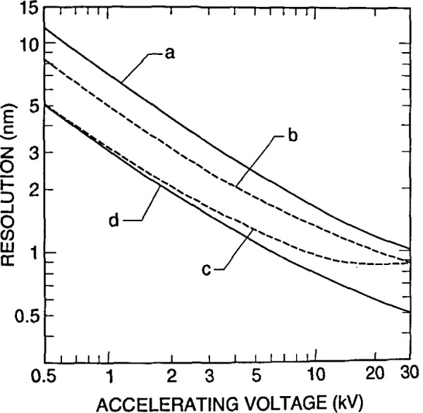
Graphical comparison of the calculated resolution capabilities of different designs and applications of field emission scanning electron microscopes. (a) Standard “pinhole” type final lens with the secondary electron detector in the “normal” location within the specimen chamber; (b) extended-field lens with the electron detector in the “normal” location; (c) extended-field lens with the detector positioned above the lens; (d) in-lens ultra-high resolution instrument (courtesy of Hitachi Scientific Instruments).