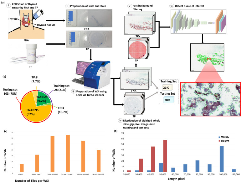
Figure 1.
Illustration of the proposed framework structure and the dataset information. (a) The proposed framework structure. (i) Collection of thyroid smear samples of patients through FNA and TP; (ii) preparation of thyroid FNA and TP slides with papanicolaou’s staining; (iii) digitalization of cytological slides at 20× objective magnification using Leica AT Turbo scanner; (iv) distribution of digitized gigapixel WSIs into separate training set (21%) and testing set (79%); (v) processing of WSIs with fast background filtering; (vi) identification of PTC tissues of individual WSIs using the proposed deep learning model in seconds. (b) Distribution of thyroid FNA and TP cytological slides for training and testing. (c) Distribution of tile numbers per WSI. (d) Size distribution of the WSIs with width and height as blue and red, respectively.

