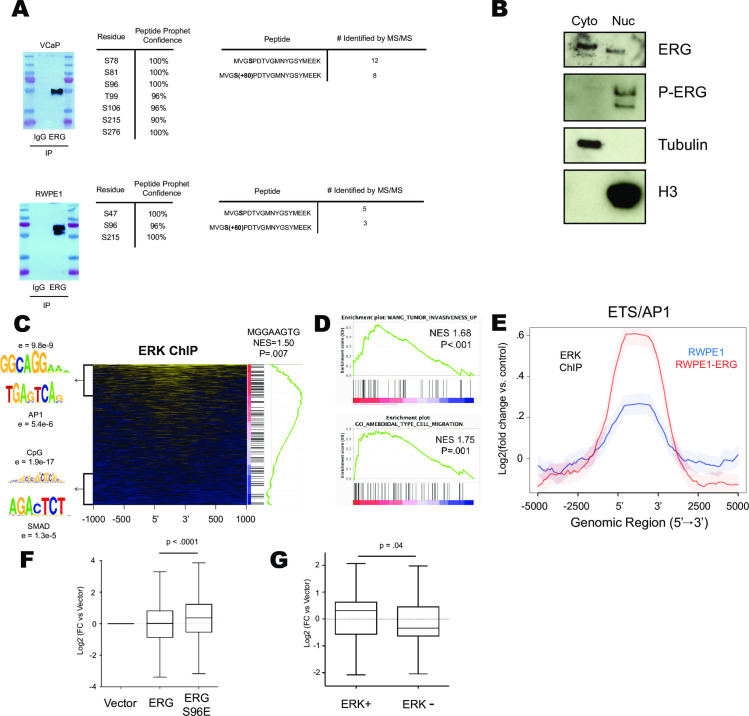
Fig 2. Ras/ERK signaling occurs at ETS/AP1 genomic locations to regulate cell migration.
(A) MS/MS analysis of ERG immunoprecipitated from VCaP and RWPE-ERG cells. Data tables show phospho-residue number of ERG NCBI Isoform 1 and compares number of apo/phospho peptides with coverage of S96. (B) Cytoplasmic/Nuclear fractionation of RWPE-ERG immunoblotted for indicated proteins. (C) Heatmap of ERK ChIP-Seq data centered on ERG bound regions [15] depicting motifs over-represented in the top 500 ERK enriched and bottom 500 ERK depleted genes and GSEA analysis of ERK enrichment across ERG bound regions run for motif analysis. (D) GSEA analysis of ERK ChIP-Seq data enrichment ranked on ERG bound regions. (E) ERK ChIP-Seq data centered on consensus ETS/AP1 motifs previously found to be ERG bound in RWPE1. (F) Log2(FC) of nearest genes bound by ERG in RWPE1 cells determined by ChIP-Seq in cells expressing ERG or ERG S96E. (G) Log2(FC) of nearest gene to ERG alone or ERG/ERK co-bound regions.

