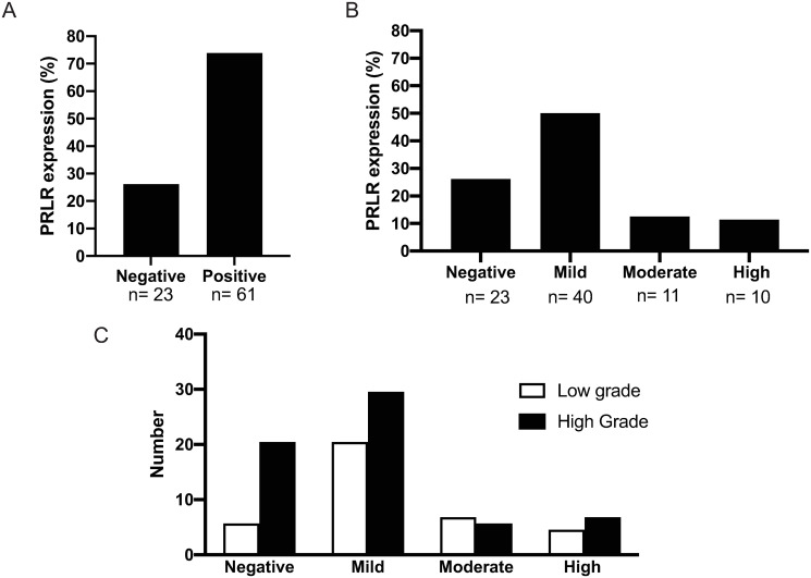
Fig 3. Prolactin receptor immuno-reactivity in ovarian cancer tissues.
Histological analyses of a tissue microarray (TMA) with 84 ovarian cancer (OC) and normal ovarian tissues as a control. A) PRLR immuno reactivity was detected in 61 out of 84 OC patients (72%) while 23 patients were negative (27%). B) PRLR staining in OC tissues; a mild level of PRLR staining (<25% of cells) was detected in 44 out of 84 patients (50%), a moderate level (25–50% of cells) was found in 11 patients (12%) and a high level (>50% of cells) was fond in 10 patients (11%). C) Percentages of PRLR positive cells were also shown in OC patients with moderate and high-grade tumour. Mild PRLR staining was detected in 30% of high-grade tumors and 20% of low-grade tumors, moderate level in 6% of high-grade tumors and 7% of low-grade tumors and high-level staining level was found in 7% of high-grade tumors and 5% of low-grade tumors. All percentages were obtained by two independent pathologist and presented with 95% confidence intervals.

