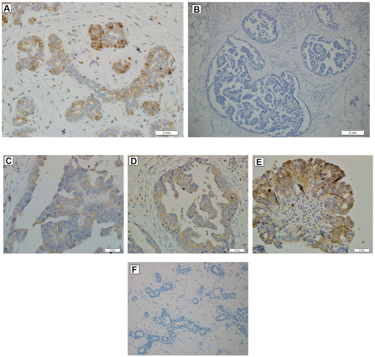
Fig 4. Immuno-histochemical staining using the PRLR antibody in ovarian cancer tissue.
(A) Breast tissue section shows moderate intensity of cytoplasmic staining with PRLR antibodies and was used as positive control. (B) OC tissue section with negative staining for PRLR expression. Grade of staining in OC tissue sections according to staining percentage, (C) Mild (<25% of the section area) level (D) Moderate (25–50% of the section area) level (E) High-grade (>50% of the section area) level. (F) Breast tissue section with negative staining for PRLR expression.

