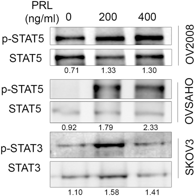
Fig 5. Prolactin signaling in ovarian cancer cells.

All cells were kept under serum free conditions overnight and later exposed to 200 and 400 ng/ml human PRL or to control PBS for 20 mints. Following protein extraction and gel electrophoresis phospho- and total STAT5, phospho- and total STAT3 were analyzed using Western blotting. A representative image selected from three independent experiments is shown.
