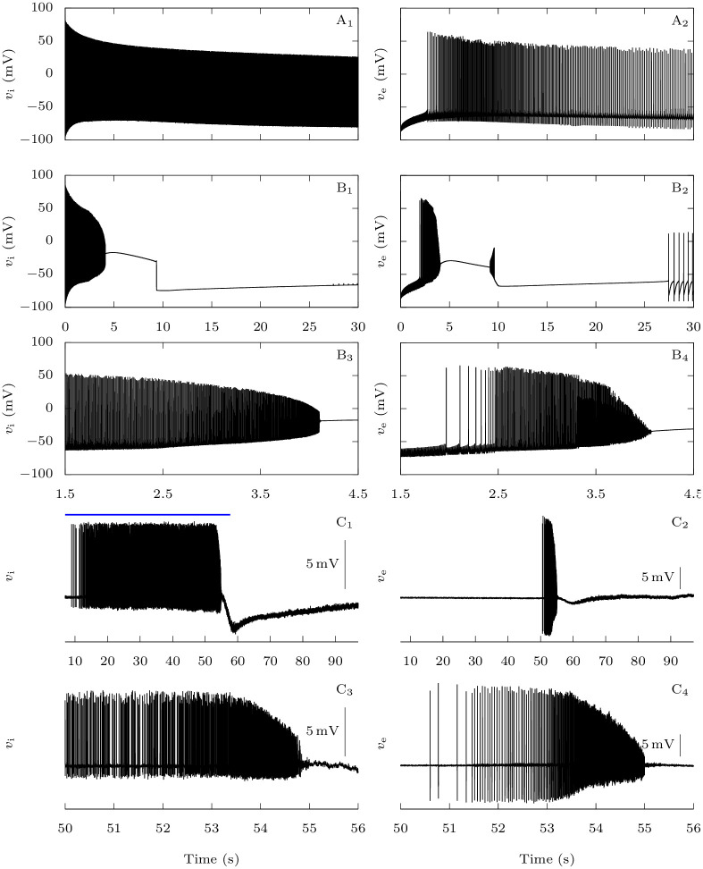
Fig 4. Effect of the FHM-3 mutation on CSD initiation: Representative time traces.
A-B: Model simulations. Starting from neurons at rest, similarly to Fig 2, we stimulated them with constant excitatory conductances gD,i = gD,e = 0.3 mS · cm−2 for 30 s. A: No persistent sodium current for the GABAergic neuron (pNa,P = 0%). B: Pathological condition (pNa,P = 15%). C: Experimental data. Representative dual juxtacellular-loose patch voltage recordings of a GABAergic interneuron (C1 and C3) and a pyramidal neuron (C2 and C4) showing the dynamics of the firing at the site of CSD initiation; CSD was induced by spatial optogenetic activation of GABAergic neurons and is the slow negative deflection observable in C1 and C2. The blue bar in C1 shows the optogenetic stimulation with blue light.

