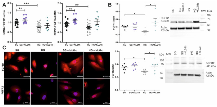
Figure 4.
Klotho increases the mRNA and protein expression of FGFRs under standard glucose conditions and recovers it after its decrease under high glucose concentration. (A) mRNA expression of the FGFR1 and FGFR2 genes increased in human podocytes after 24-h incubation with Klotho (0.5 nM; +KL24h) in the cell medium under standard glucose (SG; 11 mM; FGFR1: ** p = 0.0075, vs. SG, unpaired t-test, n = 11–19; FGFR2: ** p = 0.0038, vs. SG, unpaired t-test, n = 12–20). A trend was also found toward the rescue of FGFR1 and FGFR2 gene expression by 24-h incubation with Klotho after its initial drop under high glucose conditions (HG, 30 mM glucose, 5 days; HG+KL24h vs. HG). Detailed statistical data for the SG vs. HG comparisons are presented in Figure 3 (FGFR1: *** p = 0.0004; FGFR2: ** p = 0.0014). (B) Protein levels of FGFR1 and FGFR2 increased in human podocytes that were incubated with Klotho for 24-h (normalized to actin, HG+KL24h vs. HG: FGFR1: * p = 0.04, unpaired t-test, n = 5; FGFR2: * p = 0.03, unpaired t-test, n = 6–7; Figure 4B) after its initial drop under hyperglycemic conditions (normalized to actin, HG vs. SG: FGFR1: * p = 0.01, unpaired t-test, n = 5; FGFR2: * p = 0.03, unpaired t-test, n = 6–7; Figure 4B). (C) An increase in the protein expression of FGFR1 and FGFR2 after 24-h incubation with Klotho under both SG and HG conditions was also observed by the immunofluorescent staining of immortalized human podocytes (FGFR1-2 in red; DAPI-blue). MW, molecular weight.

