Figure 3.
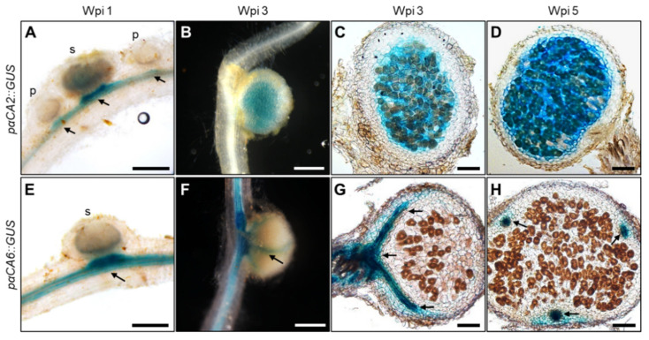
Histochemical analysis of GUS expressions driven by LjαCA2 and LjαCA6 promoters in developing root nodules. Expression patterns of GUS reporter were analyzed for pαCA2::GUS (A–D) and pαCA6::GUS (E–H). Nodules at different developmental stages were investigated, including young nodules at 1 wpi (A,E) and mature nodules at 3 wpi (B,C,F,G) and 5 wpi (D,H). pαCA2::GUS and pαCA6::GUS constructs were introduced into wild-type plants using stable transformation. T2 generation plants were used for GUS staining experiments. Images are representative of at least eight independent transgenic plants. Black arrows indicate the vascular bundle. p, primordia; s, small nodule. Scale bars, 200 μm (A,E); 1 mm (B,F); 100 μm (C,D,G,H).

