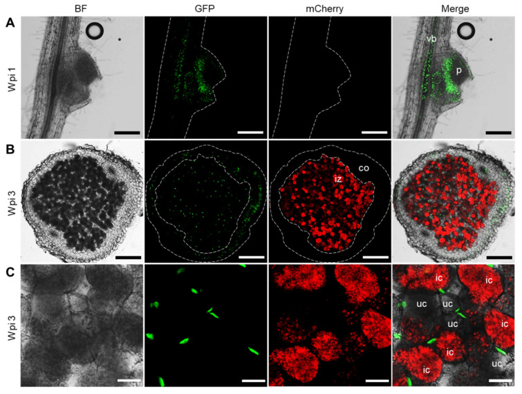
Figure 4.
Fluorescence observation of tYFP-NLS driven by LjβCA1 promoter in developing root nodule. Stable transgenic plants carrying the pβCA1::tYFP-NLS construct were inoculated with mCherry-labeled M. loti MAFF303099. Confocal microscopic images were captured at different developmental stages of root nodules, including young nodules at 1 wpi (A) and mature nodules at 3 wpi (B,C). Nuclei accumulating a green signal in the GFP channel show the fluorescence of tYFP-NLS reporter. The red signal in the mCherry channel shows the fluorescence of mCherry-labeled rhizobia. BF, bright field. Merged images of the BF, GFP, and mCherry channels were shown. vb, vascular bundle; p, primordia; iz, infected zone; co, cortical cell layers; ic, infected cell; uc, uninfected cell. Scale bars, 200 μm (A,B); 25 μm (C).

